Plan with Precision.
Proceed with Confidence.
Accomplish with Care.

Experience better accuracy and greater efficiency with uCT-guided intervention. From pre-surgical planning to follow-up evaluation, every step is designed to support confident decision-making and improve patient outcomes.

As the global population ages—especially the proportion over 60, projected to double from 12% in 2024 to 20.1% by 20501 — the demand for minimally invasive, image-guided procedures continues to rise, leading the 59% market growth of intervention from $30 billion in 2024 to estimated 43 billion in 20312.

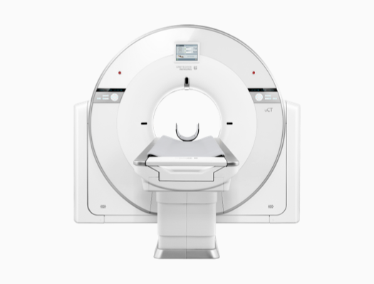
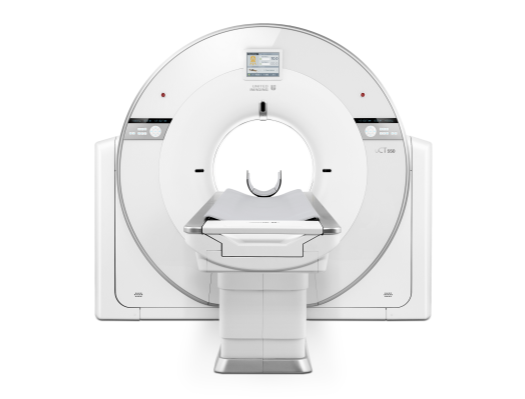
Among various interventional approaches, CT guidance offers superior spatial resolution, enabling confident execution of complex procedures such as ablation, biopsy, and drainage3. With our dedicated uCT-guided intervention solution, we strive to deliver precision at every step—so you can proceed with confidence.
1. Galan, S. "Projected World Population Distribution, by Age Group 2100." Statista, 14 Feb. 2025, www.statista.com/statistics/672546/projected-world-population-distribution-by-age-group/.
2. ReAnIn. "Interventional Radiology Market Size." Reanin.com, ReAnIn, June 2025, www.reanin.com/reports/global-interventional-radiology-market.
3. Floridi, Chiara et al. "Precision Imaging Guidance in the Era of Precision Oncology: An Update of Imaging Tools for Interventional Procedures." Journal of clinical medicine vol. 11,14 4028. 12 Jul. 2022, doi:10.3390/jcm11144028

Precision Driven

2D and 3D Path Measurement

By providing 2D and 3D path measurement from various angles, uCT Family enables a more comprehensive evaluation of the needle entry path for both routine and complicated clinical scenarios.

With pre-procedural path measurement, you can be better prepared with enhanced confidence for the following intervention treatments while maintaining low invasiveness for patients.

Flexible Scanning Modes

uCT Family could provide you with four scanning options to better adapt various clinical scenarios.
Single Axial

Axial scan for fixed coverage

Single Helical

Helical scan for broader coverage

Continuous

Continuous scan with preset intervals to reduce exposure time and dose

Fluoro

Real-time imaging

Bi-directional Gantry Tilt

30° Bi-directional physical gantry tilt allows you to identify an optimized angle with reduced amount of metal in the direct path of the beam to effectively reduce streak artifacts1.

Moreover, with the tilted angle, uCT Family can also avoid direct exposure to vulnerable organs for patients2.
1. Jones, A. Kyle, et al. "Best Practice Guidelines for CT-Guided Interventional Procedures." Journal of Vascular and Interventional Radiology, vol. 29, no. 4, Apr. 2018, pp. 518–19, https://doi.org/10.1016/j.jvir.2017.10.021.
2. Tarkiainen, Jeremias et al. "RADIATION DOSE OF THE EYE LENS IN CT EXAMINATIONS OF THE BRAIN IN CLINICAL PRACTICE-THE EFFECT OF RADIOGRAPHER TRAINING TO OPTIMISE GANTRY TILT AND SCAN LENGTH." Radiation protection dosimetry vol. 199,5 (2023): 391-398. doi:10.1093/rpd/ncad002

Intuitive Couch-side Control

Easy Operate

The couchside controller provides instinctive control you need during the operation with intuitive touch-pad.

The position memory function could support the couch return to the same position for procedure after scanning.

The window width and window level shortcut key allows you to smoothly choose the proper view for the image, streamlining the workflow.

Patient-centered

uCT Family provides low kV scanning option during intervention procedure to as low as 70 kVp. This allows you to operate under low dose condition, reducing the radiation for both you and your patients while maintaining high image quality for confident intervention operation.

uCT Family with bore size up to 82 cm*. The spacious design not only enhances patient accessibility and comfort but also provides you vast operating room to perform meticulous procedures.

*Applicable to uCT 820, uCT 868 and uCT 960+

uCT Family could provide patient table load up to 318 kg*, which can better accommodate for obese patients, further extending patient accessibility.

*Applicable to uCT 868 and uCT 960+ as standard configuration

Low kV scanning

uCT Family provides low kV scanning option during intervention procedure to as low as 70 kVp. This allows you to operate under low dose condition, reducing the radiation for both you and your patients while maintaining high image quality for confident intervention operation.

Wide Space

uCT Family with bore size up to 82 cm*. The spacious design not only enhances patient accessibility and comfort but also provides you vast operating room to perform meticulous procedures.

*Applicable to uCT 820, uCT 868 and uCT 960+

Robust Patient Table

uCT Family could provide patient table load up to 318 kg*, which can better accommodate for obese patients, further extending patient accessibility.

*Applicable to uCT 868 and uCT 960+ as standard configuration

Clinical Case

Abdominal Puncture Biopsy

The system supports both 2D and 3D path planning, enabling comprehensive visualization of the needle trajectory within or across image slices. It can automatically calculate parameters including the path length and the path angle with Y axis—facilitating precise and confident procedural planning.

  • Scan Parameters
  • Scan Modes: Single Helical
    120 kVp
    30 mAs
    CTDIvol: 2.645 mGy
    ED: 0.48 mSv
  • Reconstruction Parameters
  • Thickness/Increment:
    2.5*2.5 mm
    Kernel: B_SOFT_B
  • Scanner Model
  • uCT 820
Thoracic Fiducial Marker Placement

Fluoro mode provides real-time visualization of the needle tip, implanted markers, and target lesion, thereby offering precise positional guidance throughout the intervention.

  • Scan Parameters
  • Scan Modes: Fluoro
    120 kVp
    29 mAs
    CTDIvol: 13.999 mGy
    ED: 0.196 mSv
  • Reconstruction Parameters
  • Thickness/Increment:
    10*10 mm
    Kernel: B_SHARP_C
  • Scanner Model
  • uCT 960+
Lung Biopsy

With the help of single axial and continuous modes, the position of the needle and the lesion can be clearly displayed, providing precise guidance for interventional procedures.

  • Scan Parameters
  • Scan Modes: Single Axial, Continuous
    120 kVp
    30 mAs
    CTDIvol: 3.0 mGy
    ED: 0.1 mSv
  • Reconstruction Parameters
  • Thickness/Increment:
    5.5*5.5 mm
    Kernel: B_VSHARP_B
  • Scanner Model
  • uCT 550
Spinal Radiofrequency Ablation

With clear visualization of the needle, it provides exceptional confidence for meticulous interventional procedures.

  • Scan Parameters
  • Scan Modes: Single Helical
    120 kVp
    30 mAs
    CTDIvol: 2.913 mGy
    ED: 0.041 mSv
    Pitch: 0.6750
  • Reconstruction Parameters
  • Thickness/Increment:
    1.25*1.25 mm
    Kernel: B_SHARP_C
  • Scanner Model
  • uCT 960+

Supported uCT Family Member

uCT 960+

uCT 868

uCT 820

front view of uCT 780
uCT 780

uCT Orion Elite

uCT 550