Attainable Intelligence.
Simply Masterful.

Born with AI, uCT 960+ is an ultra-premium CT scanner equipped with industry-leading AI-empowered technologies. The unparalleled AI features gives it the capability to deliver unmatched image quality for confident diagnoses with exceptional clinical efficacy. By infusing AI with elevated hardware platform, uCT 960+ offers precise imaging and ease of use for all types of patients from routine to advanced applications, including cardiac, acute care, trauma, and oncology.

Experiencing unrestricted cardiac imaging.

True Cardiac Vision

Improving your clinical applications and efficiency through innovative AI technologies.

AI-driven Smart Imaging

Expanding clinical offerings, leaving no patients behind.

All-rounded Clinical Versatility

True Cardiac Vision

Cardiovascular diseases are the most common cause of death globally and Coronary CT Angiography (CCTA) is the examination method that is highly recommended by European Society of Cardiology (ESC), American Heart Association (AHA), and many other institutions. However, since the heart is constantly beating during the examination, cardiac imaging is one of the most challenging scenarios. uCT 960+ equipped with unparallel AI features has now made it simple as breathing. Paired with elevated hardware, 16 cm Z-axis coverage detector and 0.25 sec rotation speed, uCT 960+ can provide seamless examination experience and meanwhile, eliminates the restriction of patients, allowing high heart rate, arrhythmia, and free breathing cardiac imaging.

CardioBoost
AI-empowered cardiac imaging reconstruction.
Contrast Boost
Specialized 3D Neural Network
The 3D convolutional neural network is specially trained using well-sorted data with various cardiac anatomical structures. This helps enhancing the clarity of the interested region in cardiac imaging.
 
Up to 99%*

LCD enhancement with the same dose level

 
Up to 70%*

Dose reduction with the same LCD level


*CardioBoost images compared with FBP based on phantom tests. Data on file.

Resolution Boost
Spatial Attention Module
Bringing clearer visualization of the targeting area such as plaques and stents.
 
Up to 72%*

Improvements in spatial resolution with the same radiation dose


*CardioBoost images compared with FBP based on phantom tests. Data on file.

Artifacts Suppression
With spatial attention module, it can reduce blooming artifacts, and therefore allowing a precise evaluation about the degrees of coronary stenosis.
Improved Depiction of Non-calcified Plaques

CardioBoost delivers higher resolution of distal vessels and, meanwhile, provides clearer depiction with lower noise of the non-calcified plaques.

  • CorCTA Axial
  • kV: 100 mAs: 187
    0.25 s/rotation
    CTDIvol: 21.13 mGy
    ED: 4.73 mSv
  • Reconstruction
  • Matrix: 512 x 512
    Slice: 0.50 x 0.50 mm
    WW/WL: 800/150
    HIR:
    C_SOFT_AA, KARL: 8

    CardioBoost: 1
  • Contrast
  • 50 ml, 4.5 ml/s
Uncompromised by Stents

CardioBoost can reduce image noise and, meanwhile, improve the stent resolution with clear contours.

  • CorCTA Axial
  • kV: 120 mAs: 148
    0.25 s/rotation
    CTDIvol: 26.827 mGy
    ED: 5.26 mSv
  • Reconstruction
  • Matrix: 512 x 512
    Slice: 0.50 x 0.50 mm
    WW/WL: 2000/600
    HIR:
    C_SOFT_AA, KARL: 8

    CardioBoost: 5
  • Contrast
  • 50 ml, 4.5 ml/s
Refined Diagnostic Capability

CardioBoost has lower image noise, and clearer visualization of myocardial and valve structures, which aids in the diagnosis of structural heart disease.

  • CorCTA Axial
  • kV: 100 mAs: 106
    0.25 s/rotation
    CTDIvol: 11.67 mGy
    ED: 2.61 mSv
  • Reconstruction
  • Matrix: 512 x 512
    Slice: 0.50 x 0.50 mm
    WW/WL: 800/150
    HIR:
    C_SOFT_AA, KARL: 8

    CardioBoost: 1
  • Contrast
  • 50 ml, 4.5 ml/s

Intelligent Workflow for Cardiac Imaging

The AI-empowered cardiac workflow offers operational efficiencies that make complicated cardiac exams simple and intuitive.

Efficient Positioning

With uAI Vision, uCT 960+ enables precise patient positioning with a single-click for cardiac imaging.

Adaptive Scanning

CardioAssist
Evaluating the patient's heart rate and offering automated gating settings.
CardioAdapt
Detecting and skipping irregular heartbeats, thus avoiding rescan.

Motion Correction

CardioXphase
Intelligently selecting the global optimal phase and providing the optimal phase for individual coronary arteries.
CardioCapture
Precisely extracting the centerlines of coronary arteries, thus revealing the true vision underneath the blur.

Precise Image Reconstruction

CardioBoost
AI-empowered imaging reconstruction that is specialized in cardiac imaging.

Post-processing*

uOmnispace
Experiencing intelligent post-processing for unparalleled efficacy.

*Independent of CT; separate CE certification.


One-stop Combined CCTA Imaging for Panvascular Diseases

uCT 960+ enables a combined acquisition mode of ECG-gated and non-ECG-gated imaging with a rapid switching time of down to 2 seconds. This feature allows uCT 960+ to capture high-quality coronary images, along with cerebrovascular or peripheral vascular CTA images, using just one injection of contrast media.

AI-driven Smart Imaging
uCT 960+ leverages advanced AI to enhance both diagnostic confidence and clinical efficacy. AI-driven capabilities improve image quality by reducing noise and artifacts, providing clear, accurate images for confident decision-making. At the same time, AI streamlines workflows, automating key processes to boost clinical efficiency and reduce operator dependence, ultimately enabling faster and more reliable examination results.

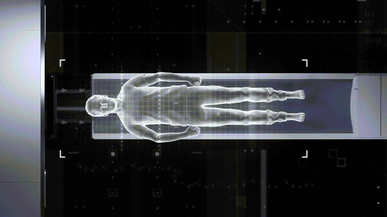
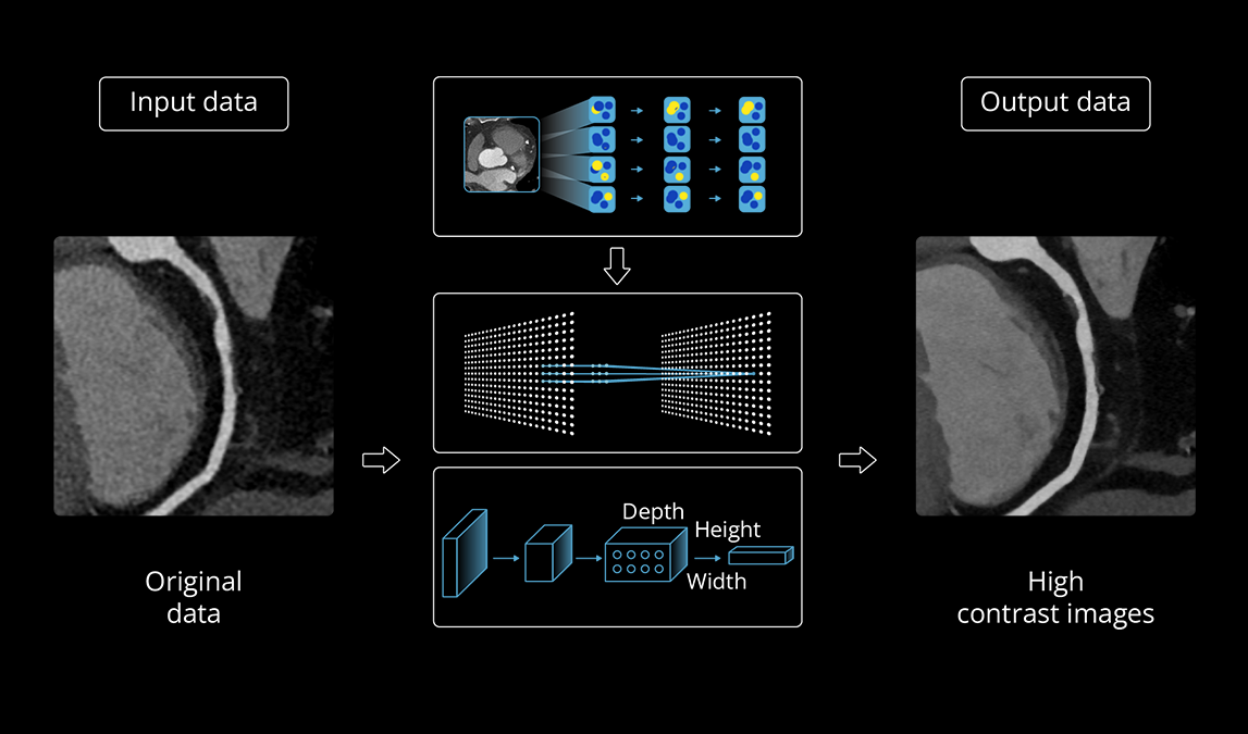
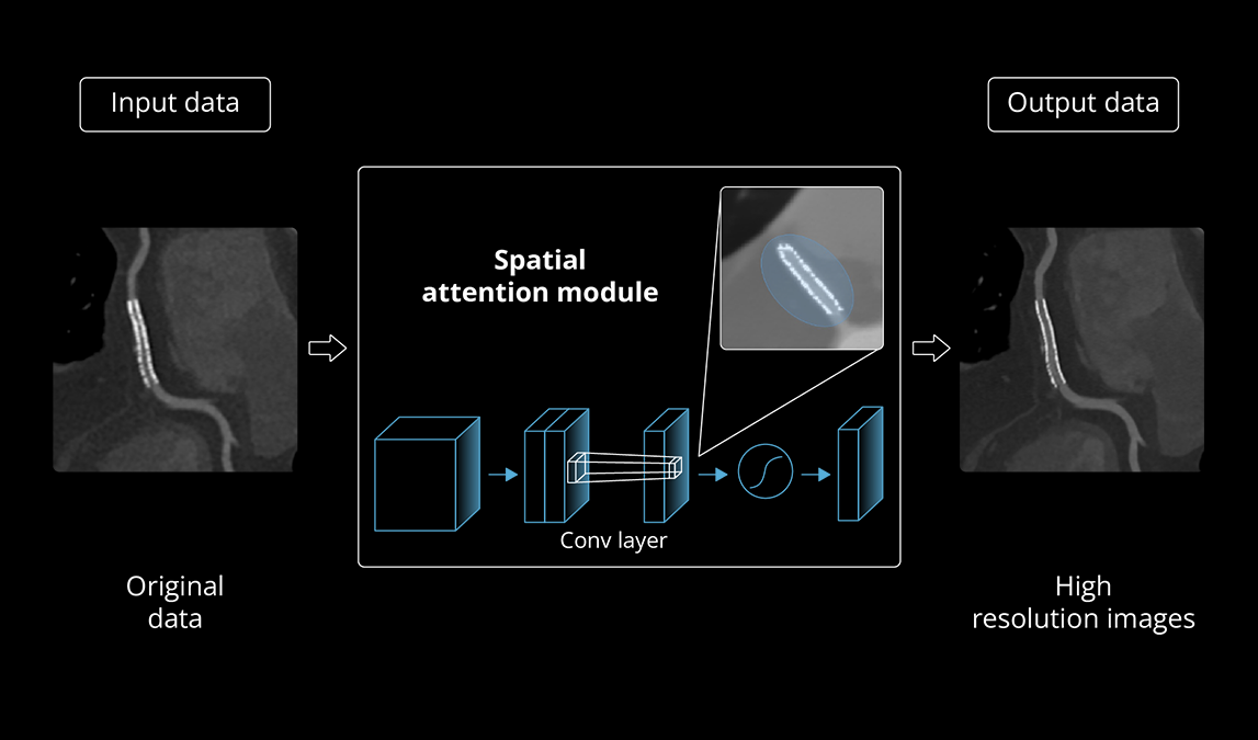
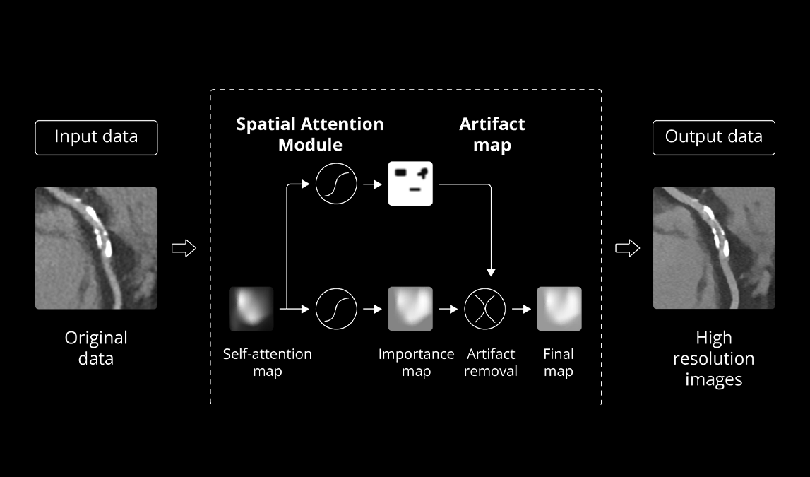
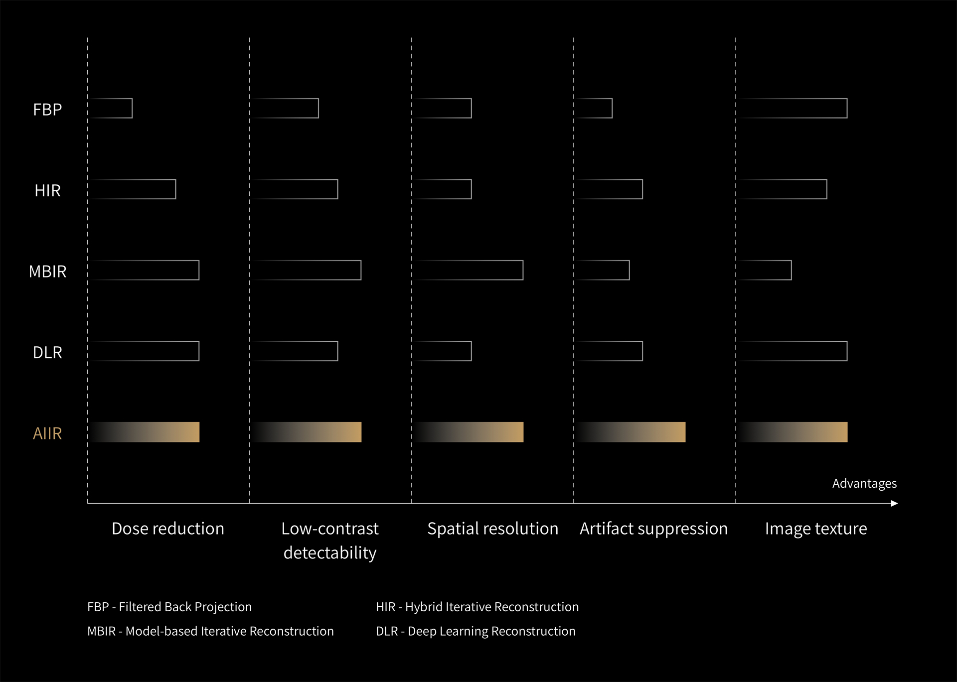
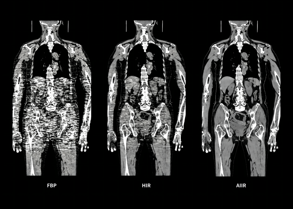
AIIR* – AI Iterative Reconstruction

As the world's first** combined AI and IR–based reconstruction, AIIR is a pioneering image reconstruction approach that integrates a holistic model-based iterative reconstruction (MBIR) with advanced deep learning technology.

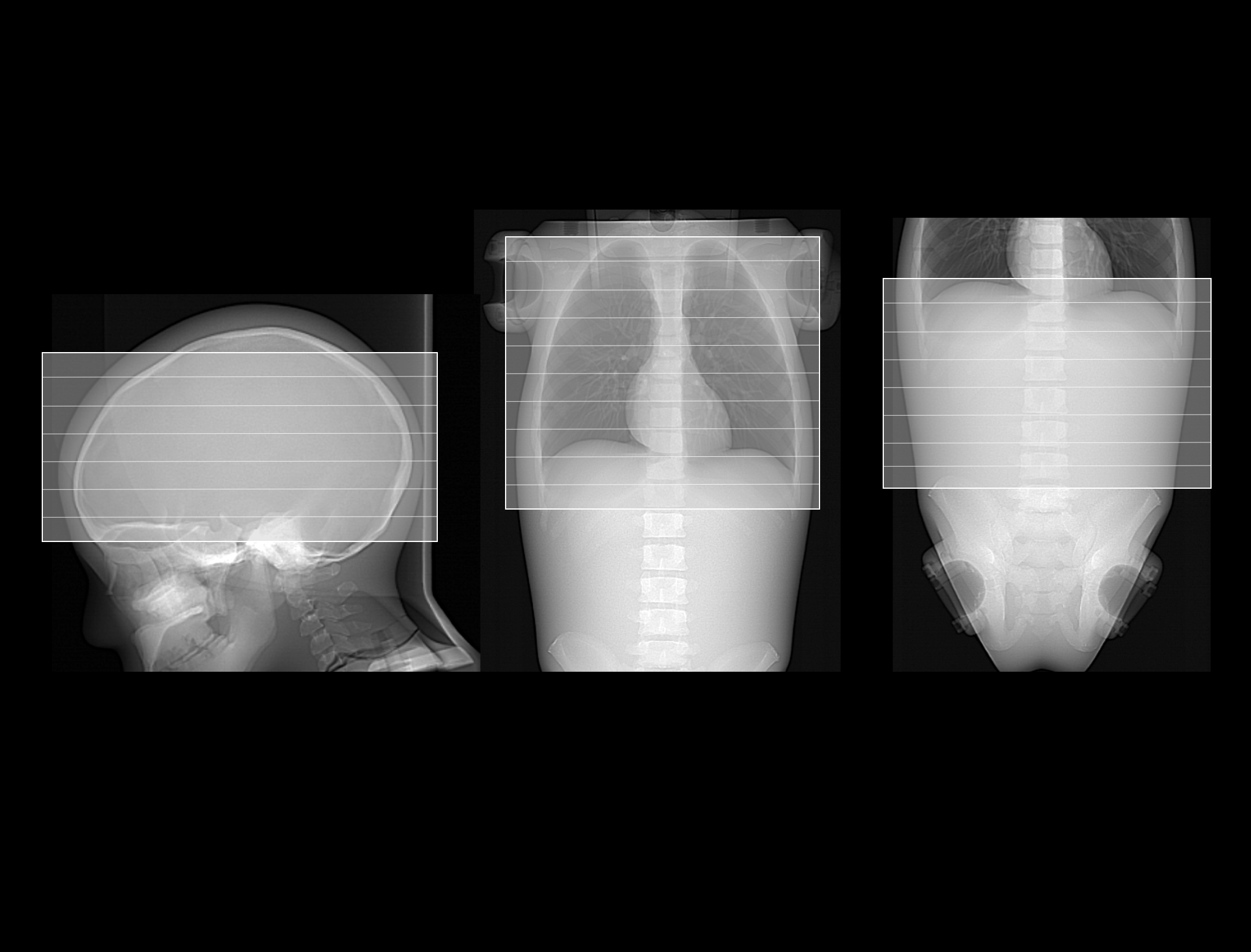
Throughout the iterative loop of forward and backward projection between the raw data domain and the image domain, AIIR consistently takes into account the accurate modeling of optics, noise, anatomy, and physics statistics. Additionally, AIIR integrates deep learning-based de-noising technology, supplanting the conventional regularization role of MBIR in the optimization reconstruction process.

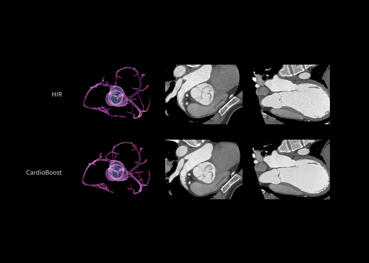
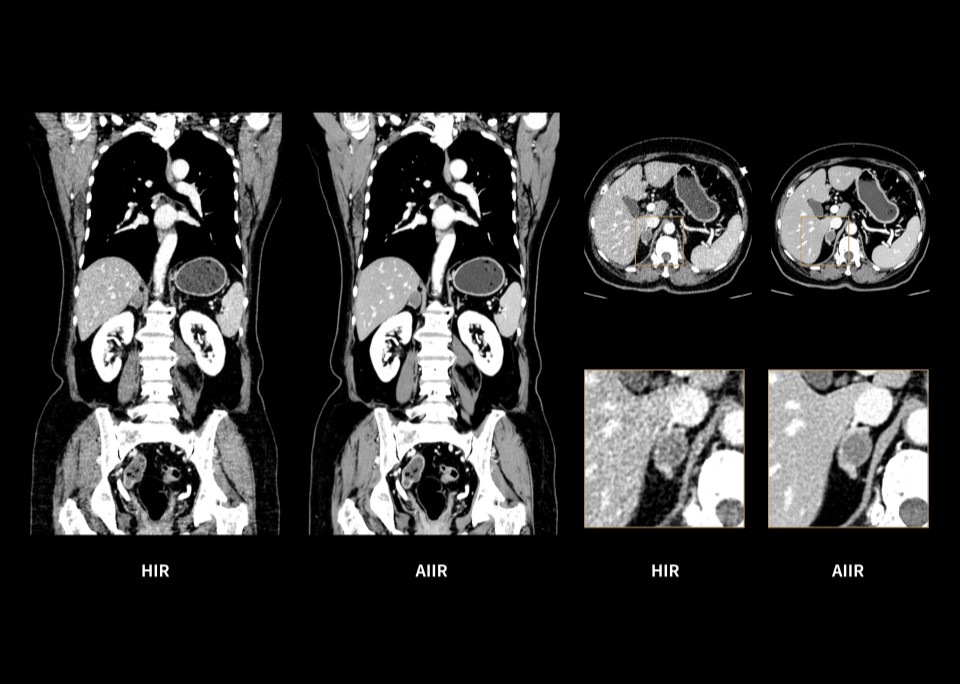
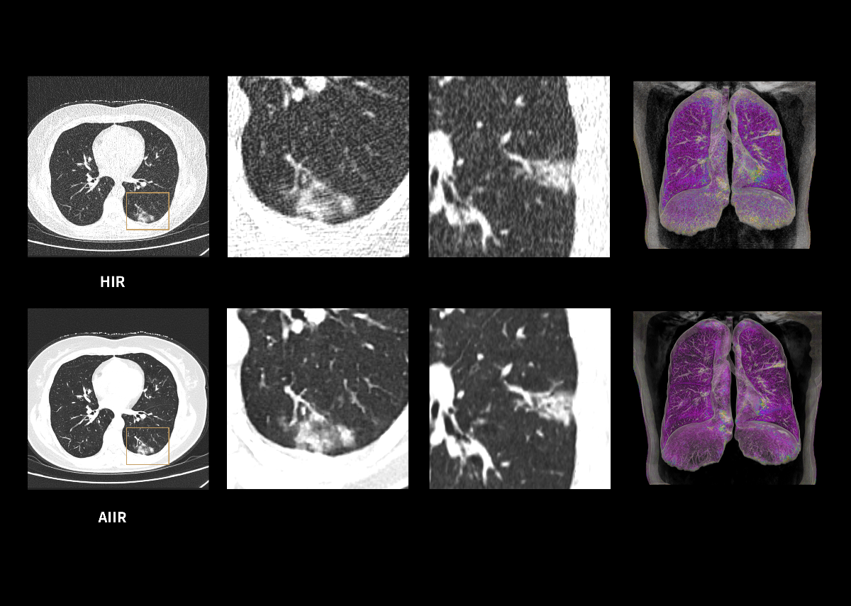
In brief, AIIR utilizes deep learning-based AI technology to attain robust noise reduction and natural image texture, while incorporating MBIR technology to achieve precise anatomical structure representation and artifact suppression. This technique surpasses the limitations of using either MBIR or deep learning reconstruction (DLR) independently.

Learn More
*Optional
**First 510(k) FDA Cleared; CE marked

Unparalleled Image Quality

With AIIR Technology, image quality is substantially improved through increased spatial resolution and low-contrast detectability (LCD), while maintaining an image texture that radiologists find familiar.
Up to 267%*

LCD improvement

Up to 150%*

Spatial resolution improvement

Up to 90%*

Dose reduction

Up to 98%*

Image noise reduction

*AIIR images compared with FBP based on phantom tests. Data on file.
High Resolution Image

Significantly reduced image noise with a 76% dose reduction*.

Enhances low-contrast detectability for clearer visualization of the right adrenal adenoma.

Suppresses artifacts in the shoulder and pelvis areas.

  • ChestUnionAbd
  • 160×0.5mm
    0.5 sec/rotation
    kV: 80;mAs: 162
    CTDIvol:3.486 mGy
    Eff. Dose: 3.48 mSv
  • Recon
  • Matrix: 512x512
    Slice: 1.0x1.0 mm
    HIR: B_SOFT_B/KARL 5
    AIIR: BodyStandard/AIIR 5
    WW/WL:300/40
  • Contrast
  • 350 mg/ml, 70 ml, 2.5 ml/s
* Kanal KM, Butler PF, Sengupta D, Bhargavan-Chatfield M, Coombs LP, Morin RL. U.S. Diagnostic Reference Levels and Achievable Doses for 10 Adult CT Examinations.
Lower Radiation Dose

The AIIR image significantly reduces image noise compared to the HIR image while greatly reducing the radiation dose.

The AIIR image shows enhanced contrast between inflammatory lesions and the lung tissue.

  • Chest Helical
  • 160×0.5mm
    0.5 sec/rotation
    kV: 80;mAs: 10
    CTDIvol:0.222 mGy
    Eff. Dose: 0.12 mSv
  • Recon
  • Matrix: 512x512
    Slice: 1.0x1.0 mm
    HIR: B_SHARP_C
    AIIR: LungStandard/AIIR:5
  • Contrast
  • -
Consistent Image Quality

For this patient who couldn't lift his arms, severe streak artifacts appeared in the FBP image.

These artifacts are effectively eliminated in the AIIR image.

  • ChestUnionAbd
  • 160×0.5mm
    0.5 sec/rotation
    Pitch: 1.37
    kV: 120 mAs: 10
    CTDIvol:0.67 mGy
    Eff. Dose: 1.6 mSv
  • Recon
  • Matrix: 512x512
    Slice: 0.5x0.5 mm
    HIR: B_SHARP_C
    AIIR: LungStandard/AIIR:5
    WW/WL:300/40
  • Contrast
  • -

uAI Vision – AI Empowered Scan Workflow

Using a 3D camera, uAI Vision enables single-click, precise, horizontal and isocenter patient positioning, allowing for easy, reproducible, and efficient scanning.
EasyPositioning

Single-click patient positioning with precise scan range planning based on the selected protocol.

EasyISO

Automatically adjusts the table height for high-quality CT images and optimized surface radiation dose distribution.

Organ-Based Auto ALARA mA
Automatically recognizing the regions of the chest and abdomen using AI-based technology, Organ-Based Auto ALARA mA enables dedicated and precise dose modulation for combined chest and abdomen scans. Compared with conventional scan modes, this innovative technology provides accurate dose modulation, consistent image quality and a streamlined clinical workflow.
A: Chest or abdomen helical protocol
An overdose or underdose may occur in the chest or abdomen area when a chest or abdomen protocol is employed to scan the combined region, as only a single set of dose modulation parameters is utilized.
B: Chest helical + abdomen helical protocols
Utilizing separate chest and abdomen protocols for scanning the combined region can increase the radiation dose due to overlapping scans from both protocols.
C: Organ-Based Auto ALARA mA
Organ-Based Auto ALARA mA allows for the implementation of a single protocol combining chest and abdomen scans, achieving equivalent image quality to two separate protocols while reducing radiation dose and scanning time.

Clinical Advantages of Organ-Based Auto ALARA mA
Based on phantom tests, the image noise in the chest and abdomen are equivalent between the Organ-Based Auto ALARA mA and the individual chest/abdomen protocols. The result of phantom tests reveal that Organ-Based Auto ALARA mA reduces radiation dose by approximately 20% due to precise dose modulation without overlapping scans.

Scan & Reconstruction – EasyRange

The EasyRange technology simplifies the workflow by automatically and precisely determining scan and reconstruction ranges through advanced deep learning techniques. This feature accommodates various routine and specialized scans, such as head, neck, chest, abdomen, spine, extremities, cardiac and combined protocols.

EasyRange for scan

In real-time, it monitors patient movement and determines the starting line and scan range.

EasyRange for reconstruction

After the scout scan, the system will automatically identify the target body part and plan the reconstruction range.

All-rounded Clinical Versatility

uCT 960+ is designed to meet the diverse needs of modern healthcare with its clinical versatility across multiple specialties. Whether it's stroke imaging, oncology, emergency care, or the unique requirements of bariatric and pediatric imaging, uCT 960+ provides reliable and high-quality results. This helps healthcare professionals deliver precise, timely diagnoses, regardless of the clinical scenario, improving overall outcomes and patient care.

Expanded Stroke Imaging Capabilities

16 cm Detector Coverage

With 16 cm z-axis detector coverage, the whole brain can be covered in a single rotation without table movement.

One-stop 4D CTA+CTP Imaging

The one-stop protocol with adjustable radiation dose and cycle time can acquire CTA and CTP information with a single contrast media injection.

Motion Freeze

Industry-first AI-based head motion correction algorithm**, which depicts the truth under motion artifacts

** First 510(k) FDA cleared; CE marked

Intelligent Stroke Evaluation

Automatically calculate critical stroke-related metrics, including cerebral blood volume (CBV), cerebral blood flow (CBF), time to peak (TTP), mean transit time (MTT), Tmax, and the mismatch ratio.

Expanded Emergency Imaging Capabilities

Single Rotation Brain Imaging

An entire scan can be completed in half a second to minimize the motion artifacts.

Up to 440 mm/s Fast Scan

Fast scans for trauma or other emergency patients.

Real Time 3D

MPR or VR preview images in real-time during the acquisition.

Intelligent and Precise Analysis

Comprehensive and accurate intelligent rib fracture analysis.

Expanded Bariatric Imaging Capabilities

82 cm Ultra-wide Bore

The expansive 82 cm bore allows for easy scanning of larger patients and provides comfortable experience for patients.

318 kg Table Load Capacity

The large load capacity, combined with an 82 cm bore size, enables efficient scanning for large patients even in challenging situations.

Ultra EFOV

AI-empowered algorithm that enables entire bore size imaging, and truncation artifacts suppression.

AIIR Technology*

The AI-based de-noising technology offers improved images with superior low-contrast detection and optimal noise texture for bariatric imaging.

*Optional

Expanded Pediatric Imaging Capabilities

16 cm Single-rotation Imaging

With a single rotation and up to 16 cm scan range, uCT 960+ can reduce sedation needs for pediatric patients.

Dedicated Pediatric Protocols

Tailored pediatric protocols, based on age and weight, can maximize efficiency and minimize radiation dose for each pediatric patients.

60 kVp Scan Mode

The 60 kVp scan mode significantly reduces radiation dose compared to higher kVp settings, while maintaining image quality.

Expanded Oncology Imaging Capabilities

Dual-Energy
Scans

Enables the acquisition of 80 kVp and 140 kVp dual-energy for spectral analysis.

16 cm Stationary Perfusion Scans

Whole organ perfusion without table movement.

Up to 40 cm
Dynamic Perfusion Scans

Dynamic perfusion scanning in a helical mode with the table moving back and forth for functional analyses.

Technical Specifications

Gantry bore size

82 cm

Gantry tilt

±30°

Detectors

16 cm Z-axis coverage 0.5 mm x 320 rows with 3D collimator grid

Number of slices

Up to 640 slices/rotation

Rotation time

Up to 0.25 sec/rotation

Temporal resolution

Up to 25 ms with CardioCapture

Table load

Up to 318 kg

kV settings

60/70/80/100/120/140 kV

Generator

Up to 100 kW