From Intelligence to Instinct
uCT 868

The uCT 868 ushers in a new era of high-end CT imaging, seamlessly integrating industry-leading AI with hardware excellence. By uniting advancements in image perception, motion detection, and workflow optimization, this cutting-edge platform empowers healthcare professionals to see more, achieve more, and operate less—instinctively unlocking a new level of diagnostic confidence and patient care.

Invisible Details, Instinctively Revealed by Intelligence


CT imaging has significantly enhanced diagnostic accuracy, yet challenges remain in achieving optimal image quality for complex cases and specialized patient populations. As the AI field evolves, deep learning is driving advancements in CT imaging by improving spatial resolution and contrast while suppressing artifacts. These innovations refine CT capabilities, making the invisible visible and ultimately improving patient outcomes.

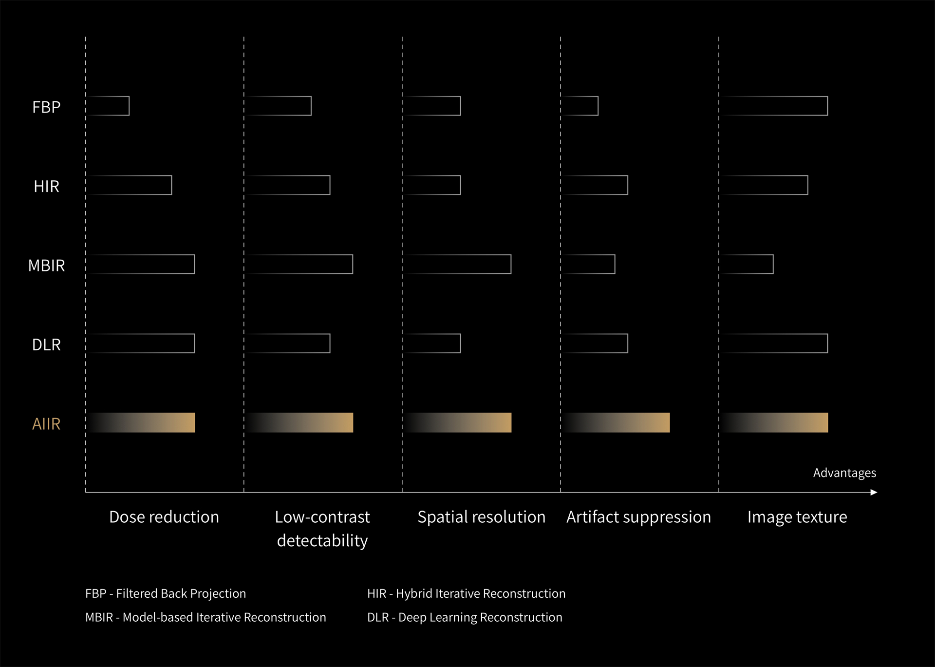
AIIR*- AI Iterative Reconstruction

Combining MBIR with deep learning leverages each method's strengths while mitigating their limitations. In AIIR, the data-fidelity term integrates system optics, detector response, and quantum noise models for each scan, preserving detailed anatomical and pathological information from raw projections. While MBIR's high regularization strength can lead to unnatural appearances—especially at low doses—AIIR replaces it with a CNN(Convolutional neural network)-based model capturing complex clinical features through millions of parameters. Together, the two technologies complement each other, allowing more precise noise differentiation and delivers improved image quality.


Learn More

AIIR* elevates image quality across all dimensions

AIIR stands out as today's most advanced CT image reconstruction architecture, setting a new standard in CT imaging with its unique design. By optimizing noise reduction, enhancing low-contrast detectability, increasing spatial resolution, and reducing artifacts, AIIR ultimately delivers exceptional image quality and superior diagnostic capabilities.
Up to 267% ※

LCD improvement

Up to 150% ※

Spatial resolution improvement

Up to 90% ※

Dose reduction

Up to 98% ※

Image noise reduction

※ AIIR images compared with FBP based on phantom tests. Data on file.
Robust Infrastructure: The Backbone of AIIR

The 0.5 mm acquisition across all FOVs and collimations unveils finer details with precision. Its fully integrated design, powered by Through-Silicon-Via (TSV) technology, revolutionizes detector architecture by reducing the signal conduction path from centimeters to micrometers, significantly lowering electronic noise and delivering ultra-low noise signal output. The 3D anti-scatter grid, precisely aligned with the X-ray source, effectively blocks scattered photons, achieving a scatter-to-primary ratio of less than 8.5%.

Equipped with high-performance GPUs, the AIIR reconstruction host meets the complex computational demands of combined deep neural networks and MBIR. Its robust infrastructure accommodates a growing volume of CT scans and large-scale data transmissions, delivering efficient imaging workflows.

High-resolution Z-Detector

The 0.5 mm acquisition across all FOVs and collimations unveils finer details with precision. Its fully integrated design, powered by Through-Silicon-Via (TSV) technology, revolutionizes detector architecture by reducing the signal conduction path from centimeters to micrometers, significantly lowering electronic noise and delivering ultra-low noise signal output. The 3D anti-scatter grid, precisely aligned with the X-ray source, effectively blocks scattered photons, achieving a scatter-to-primary ratio of less than 8.5%.

Dedicated supercomputing host*

Equipped with high-performance GPUs, the AIIR reconstruction host meets the complex computational demands of combined deep neural networks and MBIR. Its robust infrastructure accommodates a growing volume of CT scans and large-scale data transmissions, delivering efficient imaging workflows.

CardioBoost: Enhance the Diagnostic Confidence For Complicated Cardiac Imaging Through Deep Learning

CardioBoost enhances cardiac CT imaging by using 3D CNNs trained on millions of scans to overcome challenges like blooming and streak artifacts from stents, calcifications, and metallic implants. Its de-noising engine maintains low-contrast detectability, while the Spatial Attention Module sharpens resolution, reducing streaking and blooming for clearer coronary vessel and stenosis assessments.
Up to 99% ※

Low contrast detectability improvement

Up to 72% ※

Spatial resolution improvement

Up to 70% ※

Dose reduction

Up to 97% ※

Noise reduction

※ Results from phantom test
CardioBoost:Improve cardiac imaging with a unique network design

Leveraging advanced 3D CNNs trained on well-organized data for various anatomical structures, CardioBoost delivers enhanced contrast, providing clearer visualization of soft or mixed plaques.

CardioBoost incorporates a specialized module for cardiac data. The Spatial Attention Module precisely targets areas in reconstructed images, enhances spatial resolution, and enables more accurate evaluation of coronary stents.

The Spatial Attention Module reduces streaking and blooming artifacts caused by high-density structures like multiple calcium plaques at the same radiation dose, delivering sharper images.

Contrast boost

Leveraging advanced 3D CNNs trained on well-organized data for various anatomical structures, CardioBoost delivers enhanced contrast, providing clearer visualization of soft or mixed plaques.

Resolution boost

CardioBoost incorporates a specialized module for cardiac data. The Spatial Attention Module precisely targets areas in reconstructed images, enhances spatial resolution, and enables more accurate evaluation of coronary stents.

Artifact suppression

The Spatial Attention Module reduces streaking and blooming artifacts caused by high-density structures like multiple calcium plaques at the same radiation dose, delivering sharper images.

Ultra EFOV: See More Details in Extended Field of View with Deep Learning

Ultra EFOV is an innovative deep learning-based algorithm that enables full bore-size imaging, revealing additional anatomical structures and enhancing skin contours. Compared to the conventional EFOV algorithm, Ultra EFOV offers superior continuity at the edge of the Scan FOV, extends the imaging range from 50 to 82 cm, providing enhanced coverage and more detailed visualization across a broader field.

The Ultra EFOV's neural network model is designed to learn and correct different truncation artifacts within the extended field of view, resulting in better quantitative accuracy and improved image quality. This advanced algorithm provides high precision in CT density determination, with an accuracy of ±20 HU.

※ Results from phantom test

82 cm full bore-size imaging

Ultra EFOV is an innovative deep learning-based algorithm that enables full bore-size imaging, revealing additional anatomical structures and enhancing skin contours. Compared to the conventional EFOV algorithm, Ultra EFOV offers superior continuity at the edge of the Scan FOV, extends the imaging range from 50 to 82 cm, providing enhanced coverage and more detailed visualization across a broader field.

±20 HU※ quantitative accuracy

The Ultra EFOV's neural network model is designed to learn and correct different truncation artifacts within the extended field of view, resulting in better quantitative accuracy and improved image quality. This advanced algorithm provides high precision in CT density determination, with an accuracy of ±20 HU.

※ Results from phantom test

Motion Artifacts, Instinctively Suppressed by Intelligence


Motion has long been a challenge in CT imaging, often causing artifacts and reducing image clarity. This is particularly critical in dynamic areas like cardiac imaging and when patients struggle to keep their heads still during scans. Advances in technology, including improved temporal resolution and deep learning algorithms, are helping to address these issues, significantly enhancing image quality and diagnostic accuracy.

Synergy of Superior Rotation Speed and Deep Learning

Addressing motion artifacts requires combining improved temporal resolution with deep learning algorithms. While hardware improvements reduce artifacts, deep learning selects optimal phases and corrects severe motion artifacts beyond hardware's limits. This enables the uCT 868 to "freeze" motion and deliver undistorted images with enhanced coronary clarity.

In CT imaging, high acquisition speed minimizes motion artifacts and reduces radiation exposure. The native temporal resolution, which is determined by the rotation speed, directly impacts the quality of the raw data acquired. Currently, it is considered the most relevant indicator for eliminating motion artifacts in coronary artery imaging.

Greatly Diminish Motion-induced Artifacts

Addressing motion artifacts requires combining improved temporal resolution with deep learning algorithms. While hardware improvements reduce artifacts, deep learning selects optimal phases and corrects severe motion artifacts beyond hardware's limits. This enables the uCT 868 to "freeze" motion and deliver undistorted images with enhanced coronary clarity.

0.25 sec/r rotation speed

In CT imaging, high acquisition speed minimizes motion artifacts and reduces radiation exposure. The native temporal resolution, which is determined by the rotation speed, directly impacts the quality of the raw data acquired. Currently, it is considered the most relevant indicator for eliminating motion artifacts in coronary artery imaging.

CardioXphase: Finding the Optimal Phase for Coronary Arteries, Rather Than the Heart

Conventional methods prioritize minimal heart motion but may not correspond to the phase that provides the best image quality for coronary arteries. Instead of using just CT value, CardioXphase leverages AI to accurately extract arteries, focusing on shape regularity and edge sharpness to select the best phase for imaging, enhancing further analysis.

Different cardiac cycles have different optimal phases.
For CT systems with the detector coverage less than 16cm, data acquisition of the whole heart requires multiple cardiac cycles. This implies that each cardiac cycle will have a corresponding optimal phase, especially in the case of irregular heart rhythms. CardioXphase chooses the optimal phase independently from each cardiac cycle, improving the overall coronary artery image quality.

Intelligent and thorough evaluation

Conventional methods prioritize minimal heart motion but may not correspond to the phase that provides the best image quality for coronary arteries. Instead of using just CT value, CardioXphase leverages AI to accurately extract arteries, focusing on shape regularity and edge sharpness to select the best phase for imaging, enhancing further analysis.

Dynamically adapt to each cycle

Different cardiac cycles have different optimal phases.
For CT systems with the detector coverage less than 16cm, data acquisition of the whole heart requires multiple cardiac cycles. This implies that each cardiac cycle will have a corresponding optimal phase, especially in the case of irregular heart rhythms. CardioXphase chooses the optimal phase independently from each cardiac cycle, improving the overall coronary artery image quality.

CardioCapture: Deep Learning Motion Artifact Correction Overcoming Heart Rate Limitations

With the AI-based coronary artery motion correction technology, our CT scanner is able to break the limit of system native temporal resolution and achieve an effective temporal resolution of 25ms, which greatly enhances the success rate and image clarity of coronary CTA imaging.

Conventional vessel extraction methods typically rely on CT value thresholds and fixed coronary models, which often fail, particularly when dealing with vessels affected by motion artifacts. In contrast, CardioCapture excels at accurately extracting the centerlines of various types of coronary arteries, even in challenging cases involving poor vessel quality or distal vessels.

Improve the effective temporal resolution to 25ms

With the AI-based coronary artery motion correction technology, our CT scanner is able to break the limit of system native temporal resolution and achieve an effective temporal resolution of 25ms, which greatly enhances the success rate and image clarity of coronary CTA imaging.

Capture more details with precise AI extraction

Conventional vessel extraction methods typically rely on CT value thresholds and fixed coronary models, which often fail, particularly when dealing with vessels affected by motion artifacts. In contrast, CardioCapture excels at accurately extracting the centerlines of various types of coronary arteries, even in challenging cases involving poor vessel quality or distal vessels.

Motion Freeze: AI–Powered Head Motion Artifact Elimination for Clearer Brain Structures

Motion Freeze effectively suppresses head motion artifacts, providing a clear view of brain structures without obscuring lesions. By minimizing the need for repeated scans, it saves time, conserves resources, and avoids unnecessary radiation exposure for patients.

Patient head movement typically involves multiple motion patterns, and no current algorithm can effectively eliminate the resulting artifacts. To establish a gold-standard dataset, the Motion Freeze algorithm simulates artifacts in the X, Y, and Z directions, including rotation, translation, oscillation, and mixed scenarios. By introducing diverse motion artifacts, this approach enables the trained network model to handle a broad spectrum of motion conditions.

Head motion artifacts suppression

Motion Freeze effectively suppresses head motion artifacts, providing a clear view of brain structures without obscuring lesions. By minimizing the need for repeated scans, it saves time, conserves resources, and avoids unnecessary radiation exposure for patients.

3D modeling of the motion pattern to restore the real clinical situation

Patient head movement typically involves multiple motion patterns, and no current algorithm can effectively eliminate the resulting artifacts. To establish a gold-standard dataset, the Motion Freeze algorithm simulates artifacts in the X, Y, and Z directions, including rotation, translation, oscillation, and mixed scenarios. By introducing diverse motion artifacts, this approach enables the trained network model to handle a broad spectrum of motion conditions.

Innate Efficiency, Instinctively Driven by Intelligence


Efficiency is crucial in daily scanning operations. In high-end CT systems, demands for both speed and image quality extend beyond routine applications to more challenging scenarios. By harnessing advanced hardware—such as an instant-response X-ray tube for minimal preparation time and a high-speed table for fast scanning modes—paired with intelligent technologies, uCT 868 significantly enhances workflow efficiency. This integrated approach maximizes both productivity and patient benefits, even in complex clinical settings.

Maximize Rapid, Consistent Scanning with Advanced Technologies

The tube's liquid-bearing, low-friction design enable continuous anode rotation at high speed throughout the day without stopping, accelerating workflow and boosting productivity. With little preparation time required between scans, this setup is especially beneficial in time-critical scenarios.

The uAI Vision automates ISO-center positioning, enhancing efficiency, image consistency, and reducing radiation. Additionally, Easy Range optimizes workflow by recommending personalized scan ranges based on protocols.

The Organ-based Auto ALARA mA automatically recognizes the coordinate regions of the chest and abdomen to optimize dose modulation parameters for different organs.

With an 8 cm detector width and a high table speed of up to 440 mm/s, the system enables rapid scanning, completing whole-body imaging in just a few seconds.

34 MHU tube: Instant response with minimal preparation time

The tube's liquid-bearing, low-friction design enable continuous anode rotation at high speed throughout the day without stopping, accelerating workflow and boosting productivity. With little preparation time required between scans, this setup is especially beneficial in time-critical scenarios.

AI-powered iso-center positioning and scan range planning

The uAI Vision automates ISO-center positioning, enhancing efficiency, image consistency, and reducing radiation. Additionally, Easy Range optimizes workflow by recommending personalized scan ranges based on protocols.

AI-powered dose modulation

The Organ-based Auto ALARA mA automatically recognizes the coordinate regions of the chest and abdomen to optimize dose modulation parameters for different organs.

High speed scanning capability

With an 8 cm detector width and a high table speed of up to 440 mm/s, the system enables rapid scanning, completing whole-body imaging in just a few seconds.

uOmnispace**: Transforming Productivity with Intelligent Analysis and Workflow Solutions

uOmnispace enhances imaging with automated pre-processing and pre-fetching for immediate operation. Intelligent algorithms for segmentation, extraction, and labeling reduce manual effort, while a customizable layout tailors workflows to specific imaging needs, cutting processing time and ensuring precise, consistent results.

uOmnispace provides a comprehensive suite of 3D imaging and advanced visualization tools, supporting diverse clinical needs across specialties like general radiology, oncology, cardiology, and neurology. It empowers clinicians with greater confidence in decision-making.

Hyper Realistic Rendering (HRR) transforms medical imaging data into highly detailed and lifelike 3D digital visuals. Compared to conventional VR rendering, HRR provides a more accurate and realistic depiction of medical image details and features.

Automated workflow for rapid decision making

uOmnispace enhances imaging with automated pre-processing and pre-fetching for immediate operation. Intelligent algorithms for segmentation, extraction, and labeling reduce manual effort, while a customizable layout tailors workflows to specific imaging needs, cutting processing time and ensuring precise, consistent results.

Streamlining medical diagnostics with comprehensive clinical solutions

uOmnispace provides a comprehensive suite of 3D imaging and advanced visualization tools, supporting diverse clinical needs across specialties like general radiology, oncology, cardiology, and neurology. It empowers clinicians with greater confidence in decision-making.

Elevating medical imaging with advanced 3D visualization

Hyper Realistic Rendering (HRR) transforms medical imaging data into highly detailed and lifelike 3D digital visuals. Compared to conventional VR rendering, HRR provides a more accurate and realistic depiction of medical image details and features.

** Independent of CT, separate CE certification.
Technical Specifications

Gantry bore size

82 cm

Gantry tilt

±30°

Detectors

8 cm Z-axis coverage 0.5 mm x 160 rows with 3D collimator grid

Number of slices

Up to 320 slices/rotation

Rotation time

Up to 0.25 sec/rotation

Temporal resolution

Up to 25 ms with CardioCapture

Table load

Up to 318 kg

kV settings

60/70/80/100/120/140 kV

Generator

Up to 100 kW

*Optional
**Independent of CT, separate CE certification.
***uCT 868 may not be available in all countries. Please check with your local United Imaging Healthcare representative for availability.
The uCT 868 currently supports the following label languages: English, French, German, Finnish, Italian, Polish, Portuguese, et al.
The supported manual languages include: English, Russian, Spanish, Italian, Polish, German, Greek, Romanian, Hungarian, and Brazilian Portuguese, et al.
Please contact your local United Imaging Healthcare organization for further details.