Digital. Attainable.

uMI 550 makes the state-of-art digital PET technology attainable to more customers. With the unique Integrated-Light-Guide Digital PET detector, uMI 550 provides 2.9mm NEMA resolution. The 80-slice CT system with Z-Detector enables diagnostic CT images with high spatial resolution and improved low contrast detectability.

SiPM based Integrated-Light-Guide Digital PET Detector

80-slice CT with Innovative Z-detector

Intuitive Workflow for Efficiency and Personalized Care

exploded-view image of uMI 550

Integrated-Light-Guide Digital PET Detector

 the Integrated-Light-Guide, improving light collection efficiency

Integrated-Light-Guide

Integrated-Light-Guide improves light collection efficiency and spatial resolution to achieve exceptional image quality.

black chips with SiPM technology assembled on the digital PET detector

SiPM

SiPM technology increases photoelectric conversion efficiency to reduce signal loss, yielding outstanding image quality.

numerous super-fine crystals assembled on the digital PET detector

High-precision Crystal

The ultra-fine crystal size significantly enhances image resolution.

numerous modules align on the digital PET detector

Modular Design

High-level modular design achieves remarkable system reliablilty and serviceability.

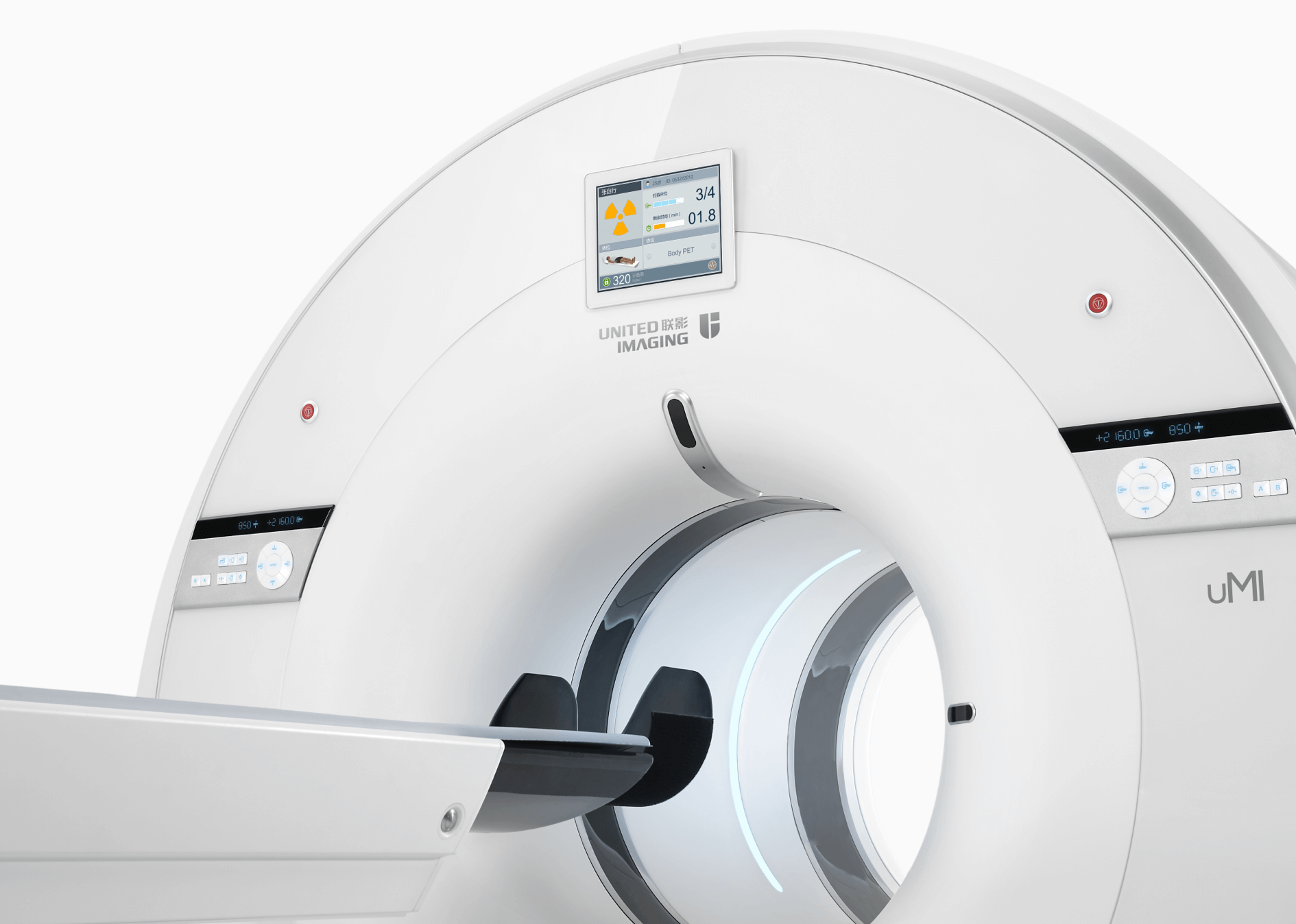
80-slice CT with Z-Detector

uMI 550's Z-Detector with ultra-low noise design and high-performance material
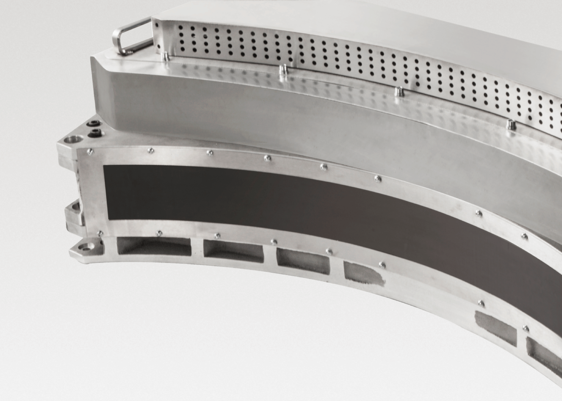
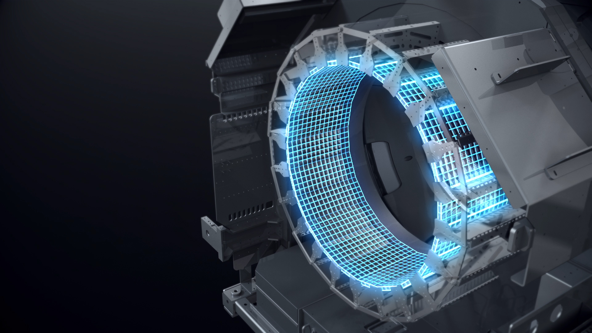
Ultra-low Noise Design

With highly integrated architecture, Z-Detector not only achieves overall noise reduction, but also helps to ensure high image quality with low radiation dose.

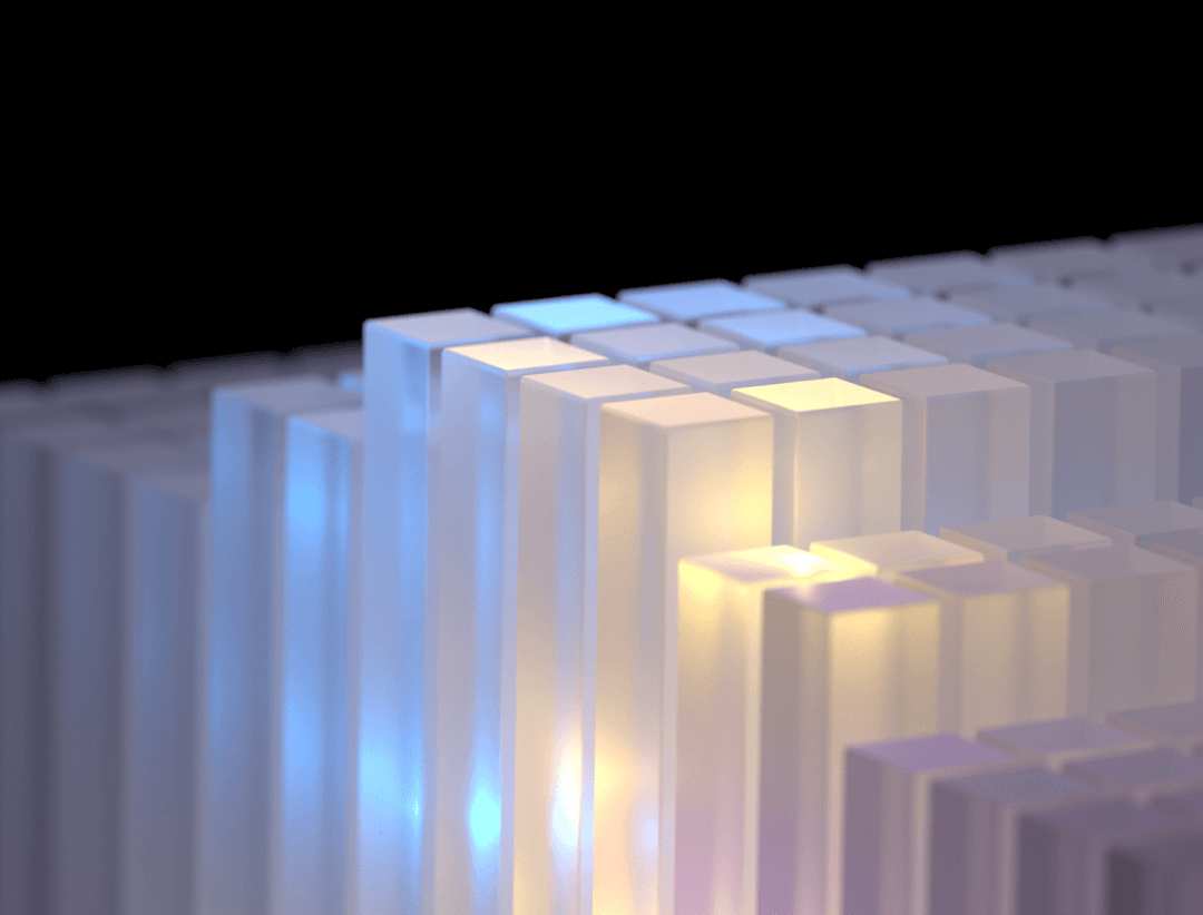
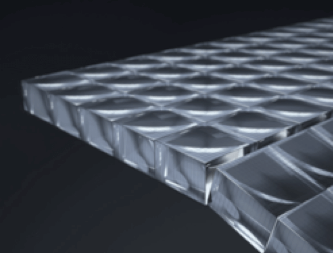
High-performance Material

The high conversion efficiency of GOS boosts X-ray conversion rate while maintaining quick response time.
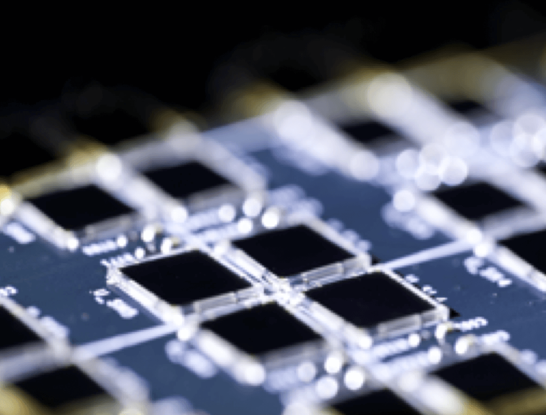
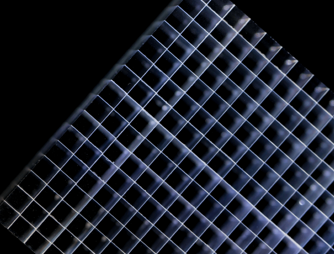
Fine Sampling

The very fine sampling voxels acquire massive information, bringing exquisite visualization of fine anatomic details.

uMI 550's Z-Detector with ultra-low noise design and high-performance material

Clarity for Imaging

A contrast of brain imaging between Conventional PET/CT and uMI 550

Ultra-High Resolution

Point-spread-function (PSF) iterative reconstruction technique helps to achieve an effective spatial resolution of 1.4mm, improving quantitative accuracy and enhancing small lesion detectability.

imaging with improved contrast and reduced noise ratio

Improved Contrast-to-Noise Ratio

With precise localization of annihilation photons, the time-of-flight (TOF) technology accelerates the image convergence with better contrast and significantly reduced image noise.

uMI 550 performing a 4-bed-whole-body scanning

Fast scanning

Large axial FOV (24cm) and high system sensitivity enable a whole-body scan in 4 bed positions.

whole-body-scan showing low dose and high sensitivity

Low Dose

High sensitivity boosts data acquisition and enables low-dose PET scans.

Intuitive Workflow for Efficiency

uMI 550 with a united-imaging logo and control panel
Automatic Quality Control Workflow

No radioactive source is required for automatic daily QC, reducing radiation exposure to the operator. Periodic full QC supports both liquid 18F and solid 68Ge sources with lower storage condition requirements. A fast NEMA tool makes the workflow convenient and efficient.

Head Motion Correction

Without the requirement for external devices, the data-driven motion correction algorithm detects motion from list-mode data during patient scans and corrects motion blurring accordingly.

Intelligent Motion Artifact Reduction

Without an external gating device, the intelligent digital self-gating technology automatically detects respiratory motions. MAC helps reduce metal artifacts and keep the important surrounding structural details.

Integrated Oncology Application

The uMI 550 offers a comprehensive solution package for tumor diagnosis and post-therapeutic evaluation on the operation console.

uMI 550 with a united-imaging logo and control panel

Clinical Case Studies

imaging of Esophagus Cancer showing different colors
Esophagus Cancer
imaging of Lymphoma with colors in white, red and blue
Lymphoma
imaging for Rectal Cancer with colors in white, red and blue
Rectal Cancer
imaging for lung cancer segmenting and analyzing the separately obtained area
Lung Cancer
12 pieces of intercept and slope brain imaging
Brain
16 pieces of Cardiac Viability Imaging with motions
Cardiac Viability Imaging
United Imaging Healthcare makes no claims about the use of the referenced radiopharmaceuticals.