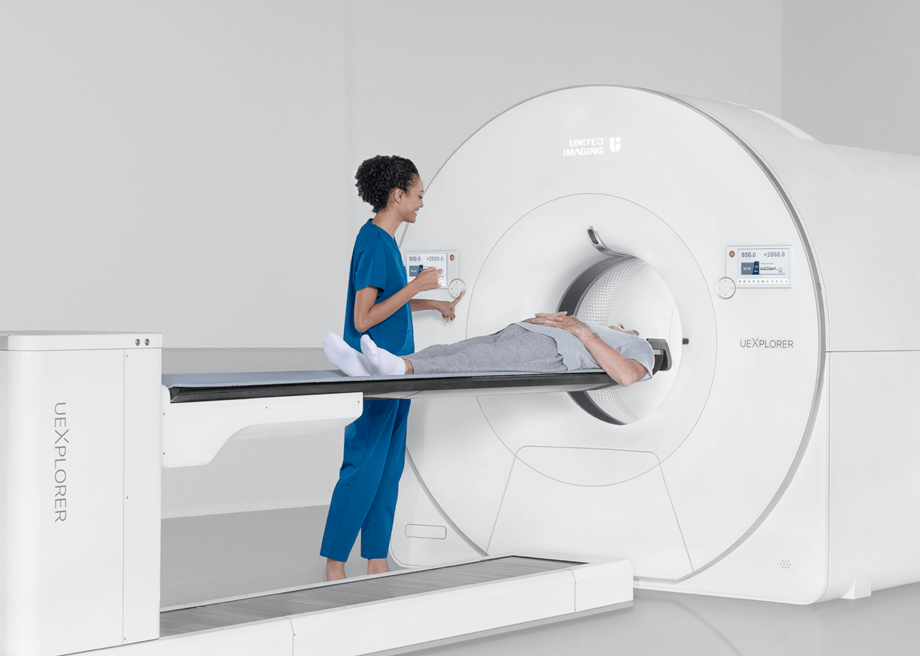
Born to Explore

The uEXPLORER is an ultra-high-resolution digital PET/CT with a 194cm axial PET field of view (FOV) that enables the entire body to be scanned in one bed position. The system offers total-body dynamic scanning, which enables ultra-low patient doses and produces ultra-high image resolution, changing the way whole-body PET/CT imaging has traditionally been performed. With total-body coverage and unprecedented sensitivity, uEXPLORER is able to capture dynamic changes to radiotracer distribution with ultra-high temporal resolution. Take healthcare applications and scientific research to the next level with uEXPLORER.

Meet the uEXPLORER

194 cm Axial FOV

Ultra-large coverage by 564,480 crystal elements

176 cps/kBq System Sensitivity*

Unrivaled sensitivity for superior image quality

*Measured with a 70 cm NEMA NU-2 phantom

2.9 mm NEMA Spatial Resolution

Functional images with anatomical details

91 Billion LORs

100x more computation power than conventional PET/CT**

**ln comparison to PET/CT with 4mm resolution and 20cm Axial FOV

lnnovation

564,480 crystal elements with unit-based design and gapless assembly 

194cm Axial FOV

564,480 crystal elements, unit-based design and gapless assembly achieve total-body coverage and minimize accumulative system error, bringing high spatial resolution and precisely fused images.

high and uniform sensitivity area of uEXPLORER

40 x Sensitivity Improvement

With 194 cm axial FOV, uEXPLORER is able to capture orders of magnitude more γ photons. The innovative cross coincidence technology enables signal acquisition across adjacent units, which gives 30 to 40-fold overall sensitivity boost over conventional PET/CT systems. Ultra-high and uniform sensitivity can now be achieved with the total-body coverage, covering all vital human organs.

integrated-light-guide digital detector with ultra-fine microscopic crystals

Integrated-Light-Guide Digital Detector

SiPM technology increases photoelectric conversion efficiency to reduce signal loss, yielding vast improvements in image quality. Ultra fine microscopic crystals significantly enhance image resolution.

fast data processing for image reconstruction

Fast Data Processing for Image Reconstruction

uEXPLORER applies a state-of-the-art hardware platform in combination with parallel data processing architecture for fast and high quality image reconstruction.

Benefits

functional images with anatomical details

Functional lmages with Anatomical Details

Ultra-high resolution and sensitivity.
High count statistics enables small voxel reconstriction.

fast total-body imaging of the thyroid nodule with high uptake

Fast Total-body lmaging

The thyroid nodule with high uptake is clearly visible on scan as short as 30 seconds. The fast imaging improves patient comfort and reduces motion artifacts.

imaging with reduced tracer dose

lmaging with Reduced Tracer Dose

0.2mCi tracer for a 100kg patient. Much lower dose (0.002mCi/kg) than the typical dose (0.08~0.15mCi/kg).

imaging tracer distribution from 90-minute post injection to 12 hours

lmaging Tracer Distribution for Longer

Scanned at up to 6.5 half lives post injection.Potential to observe radiopharmaceutical distribution over an extended period of time.

total-body dynamic imaging in one single bed

Total-body Dynamic lmaging in One Single Bed

Clear visualization of tracer distribution throughout the entire body including blood/organs/tissues over time.

Possibilities

numerous crystal elements of uEXPLORER
lmproved Lesion Detection

Improved lesion detection of tumor micro-metastases

Cell Tracking & Function

Efficacy evaluation of stem cell therapies and cancer immunotherapies*

New Pharmaceuticals Development

Early-phase total-body bio-distribution, pharmacokinetics and low-dose evaluation*

Multisystem Disease Research

Research of brain-body or multi-organ disease, such as brain-gut conditions, metabolic syndrome and endocrine homeostasis*

numerous crystal elements of uEXPLORER

Automated Quality Control

A patient is experiencing uExplorer with a staff pushing the panel.
Source-free Automatic Daily Quality Control

Automatically scans for background count rate, voltage power supply, temperature, and humidity.
Single-click for CT detector channel check and air calibration.
Reduces staff's exposure to radiation sources.

Weekly Full Quality Control with Easy-to-Handle Process

Supports both liquid 18F-FDG and solid 68Ge sources.
Lower storage condition requirement for sources.
Thorough calibration procedures help ensure accurate and consistent clinical performance over time.

Easy-to-Use NEMA Tool

Workflow-driven NEMA testing with automated analysis and report generation.
User-oriented design improves the efficiency and success rate of the workflow parameters including spatial resolution, sensitivity, noise equivalent count rate scatter fraction, accuracy and image quality.

A patient is experiencing uExplorer with a staff pushing the panel.

Focus On Patient Experience

front view of uExplorer with an illuminated patient bore
76cm PET Aperture

Improved patient comfort.

Illuminated Patient Bore

Relaxing patient experience.

3 In-bore Intercoms

Clear patient communication.

Four Touchscreen Control Panels

Control where you need it.

front view of uExplorer with an illuminated patient bore