uCT SiriuX Ultrix™ brings a new level of versatility to CT imaging. Its dual-wide coverage architecture integrates high spatial resolution, fast temporal resolution, extensive volumetric coverage, low-dose imaging, and high-definition spectral imaging within a single platform. From anatomical detail to physiological motion and material composition, it offers a broader view of what CT can reveal.
Built on a dual-wide coverage architecture, uCT SiriuX Ultrix™ delivers both ultra-fast temporal resolution and extensive volumetric coverage, unlocking new possibilities in CT performance. It precisely captures the full spectrum of physiological motion, offering a new perspective on dynamic imaging.
Powered by dual Ultrix™ detectors with 16 cm Z-axis coverage, uCT SiriuX Ultrix™ enables comprehensive dynamic imaging from brain to heart and beyond. Combined with ultra-high temporal resolution, it delivers sharper, faster, and more precise visualization of physiological motion.
Built on a dual-source CT architecture, uCT SiriuX Ultrix™ achieves enhanced physical temporal resolution for capturing rapid physiological changes with exceptional clarity. Liquid metal bearing technology ensures smooth and stable rotation, while the 34 MHU high-capacity anode supports sustained performance under demanding clinical workloads.
Twin-ring gantry design redefines CT architecture. The imaging ring integrates dual X-ray tubes and dual 16 cm wide-area detectors, while the carrier ring integrates power and thermal management for a compact, efficient design. The separable Twin-rings simplify installation and maintenance. With micron-level precision and high-speed data synchronization, uCT SiriuX Ultrix™ delivers reliable, high-precision imaging for every scan.
Powered by dual Ultrix™ detectors with 16 cm Z-axis coverage, uCT SiriuX Ultrix™ enables comprehensive dynamic imaging from brain to heart and beyond. Combined with ultra-high temporal resolution, it delivers sharper, faster, and more precise visualization of physiological motion.
Built on a dual-source CT architecture, uCT SiriuX Ultrix™ achieves enhanced physical temporal resolution for capturing rapid physiological changes with exceptional clarity. Liquid metal bearing technology ensures smooth and stable rotation, while the 34 MHU high-capacity anode supports sustained performance under demanding clinical workloads.
Twin-ring gantry design redefines CT architecture. The imaging ring integrates dual X-ray tubes and dual 16 cm wide-area detectors, while the carrier ring integrates power and thermal management for a compact, efficient design. The separable Twin-rings simplify installation and maintenance. With micron-level precision and high-speed data synchronization, uCT SiriuX Ultrix™ delivers reliable, high-precision imaging for every scan.
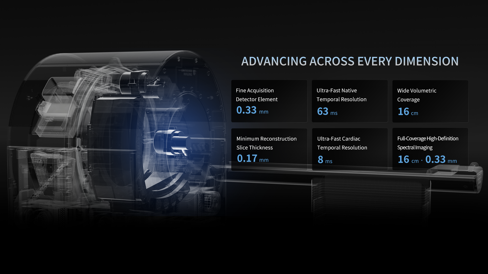
Building on a dual-source architecture, uCT SiriuX Ultrix™ adopts an advanced gantry design to overcome the challenges of increased rotational inertia and high centrifugal forces, enabling fast rotation and an ultra-high physical temporal resolution of 63 ms.
0.239 s/360°
Rotation Speed
63 ms
Native Temporal Resolution
With 2 × 16 cm wide Z-axis coverage, uCT SiriuX Ultrix™ combines ultra-fast temporal resolution with whole-organ coverage to capture physiological motion in its entirety. From cardiac motion to joint movement, complete anatomical regions can be imaged simultaneously with exceptional clarity, minimizing temporal mismatch and advancing CT from partial coverage to whole-organ synchronized imaging.
Building on a dual-source architecture, uCT SiriuX Ultrix™ adopts an advanced gantry design to overcome the challenges of increased rotational inertia and high centrifugal forces, enabling fast rotation and an ultra-high physical temporal resolution of 63 ms.
0.239 s/360°
Rotation Speed
63 ms
Native Temporal Resolution
With 2 × 16 cm wide Z-axis coverage, uCT SiriuX Ultrix™ combines ultra-fast temporal resolution with whole-organ coverage to capture physiological motion in its entirety. From cardiac motion to joint movement, complete anatomical regions can be imaged simultaneously with exceptional clarity, minimizing temporal mismatch and advancing CT from partial coverage to whole-organ synchronized imaging.
CardioCapture 2.0 achieves an effective temporal resolution of 8 ms through AI-powered cardiac motion correction. By precisely segmenting cardiac structures, tracking motion trajectories, and generating multi-phase motion fields, it enables synchronized motion correction of the coronary arteries, valves, myocardium, and ventricles for clearer visualization of cardiac dynamics throughout the entire cardiac cycle.
CardioCapture 2.0 reconstructs multi-phase images before and after the target phase, using deep learning networks to accurately extract coronary centerlines and establish inter-phase motion models. This enables precise motion correction of segmented coronary images for clearer coronary visualization.
CardioCapture 2.0 provides synchronized motion correction for the myocardium, valves, and ventricles. Powered by deep learning-based cardiac segmentation, it extracts comprehensive cardiac information, effectively suppresses motion artifacts, and significantly enhances structural clarity and diagnostic confidence.
CardioCapture 2.0 achieves an effective temporal resolution of 8 ms through AI-powered cardiac motion correction. By precisely segmenting cardiac structures, tracking motion trajectories, and generating multi-phase motion fields, it enables synchronized motion correction of the coronary arteries, valves, myocardium, and ventricles for clearer visualization of cardiac dynamics throughout the entire cardiac cycle.
CardioCapture 2.0 reconstructs multi-phase images before and after the target phase, using deep learning networks to accurately extract coronary centerlines and establish inter-phase motion models. This enables precise motion correction of segmented coronary images for clearer coronary visualization.
CardioCapture 2.0 provides synchronized motion correction for the myocardium, valves, and ventricles. Powered by deep learning-based cardiac segmentation, it extracts comprehensive cardiac information, effectively suppresses motion artifacts, and significantly enhances structural clarity and diagnostic confidence.
Powered by its advanced Ultrix detector and precision imaging technologies, uCT SiriuX Ultrix™ delivers exceptional spatial resolution for visualizing fine anatomical structures with remarkable clarity. By enhancing signal acquisition and image reconstruction, it reveals subtle details that can support confident diagnosis across a broad range of clinical applications.

The Ultrix™ detector features high-precision dicing for 0.33 mm pixel size at isocenter. A reduced reflector thickness enables an expanded active detecting area, improving photon collection efficiency for low-noise, high-resolution imaging. This supports image quality in low-dose and large-patient examinations.
0.33 mm
Pixel Size @ Iso-center
The Ultrix™ detector integrates a 3D-printed thin-wall anti-scatter grid (ASG) with one-to-one alignment between grid cells and scintillator pixels for effective scatter rejection. Made from a high-density heavy metal alloy, the ASG combines structural rigidity with a thin-wall design, supporting efficient X-ray transmission and photon detection for high SNR and clear image quality.
The Ultrix™ detector features high-precision dicing for 0.33 mm pixel size at isocenter. A reduced reflector thickness enables an expanded active detecting area, improving photon collection efficiency for low-noise, high-resolution imaging. This supports image quality in low-dose and large-patient examinations.
0.33 mm
Pixel Size @ Iso-center
The Ultrix™ detector integrates a 3D-printed thin-wall anti-scatter grid (ASG) with one-to-one alignment between grid cells and scintillator pixels for effective scatter rejection. Made from a high-density heavy metal alloy, the ASG combines structural rigidity with a thin-wall design, supporting efficient X-ray transmission and photon detection for high SNR and clear image quality.
The uCT SiriuX Ultrix™ features dual liquid-metal-bearing X-ray tubes with a minimum focal spot of 0.4 × 0.5 mm, helping reduce geometric penumbra and support high spatial resolution. Combined with dual Ultrix™ detectors, the X-ray tubes and detectors form an integrated imaging chain for detailed image visualization. Multiple focal spot options provide flexibility for high-resolution imaging and high-throughput scanning.
0.4 x 0.5 mm
Ultra-small Focal Spot Size
The uCT SiriuX Ultrix™ employs 3D flying focal spot technology, dynamically shifting the focal spot across the X, Y, and Z axes. This enables double or quadruple sampling per projection in both the transaxial and longitudinal directions, substantially increasing effective sampling density and supporting a minimum slice thickness of 0.17 mm.
0.17 mm
Minimum Reconstruction Slice Thickness
The uCT SiriuX Ultrix™ features dual liquid-metal-bearing X-ray tubes with a minimum focal spot of 0.4 × 0.5 mm, helping reduce geometric penumbra and support high spatial resolution. Combined with dual Ultrix™ detectors, the X-ray tubes and detectors form an integrated imaging chain for detailed image visualization. Multiple focal spot options provide flexibility for high-resolution imaging and high-throughput scanning.
0.4 x 0.5 mm
Ultra-small Focal Spot Size
The uCT SiriuX Ultrix™ employs 3D flying focal spot technology, dynamically shifting the focal spot across the X, Y, and Z axes. This enables double or quadruple sampling per projection in both the transaxial and longitudinal directions, substantially increasing effective sampling density and supporting a minimum slice thickness of 0.17 mm.
0.17 mm
Minimum Reconstruction Slice Thickness
uCT SiriuX Ultrix™ combines low-noise detector technology with intelligent reconstruction for high-quality imaging at low radiation dose. Its dose-efficient imaging supports a broad range of clinical applications while maintaining diagnostic image quality.
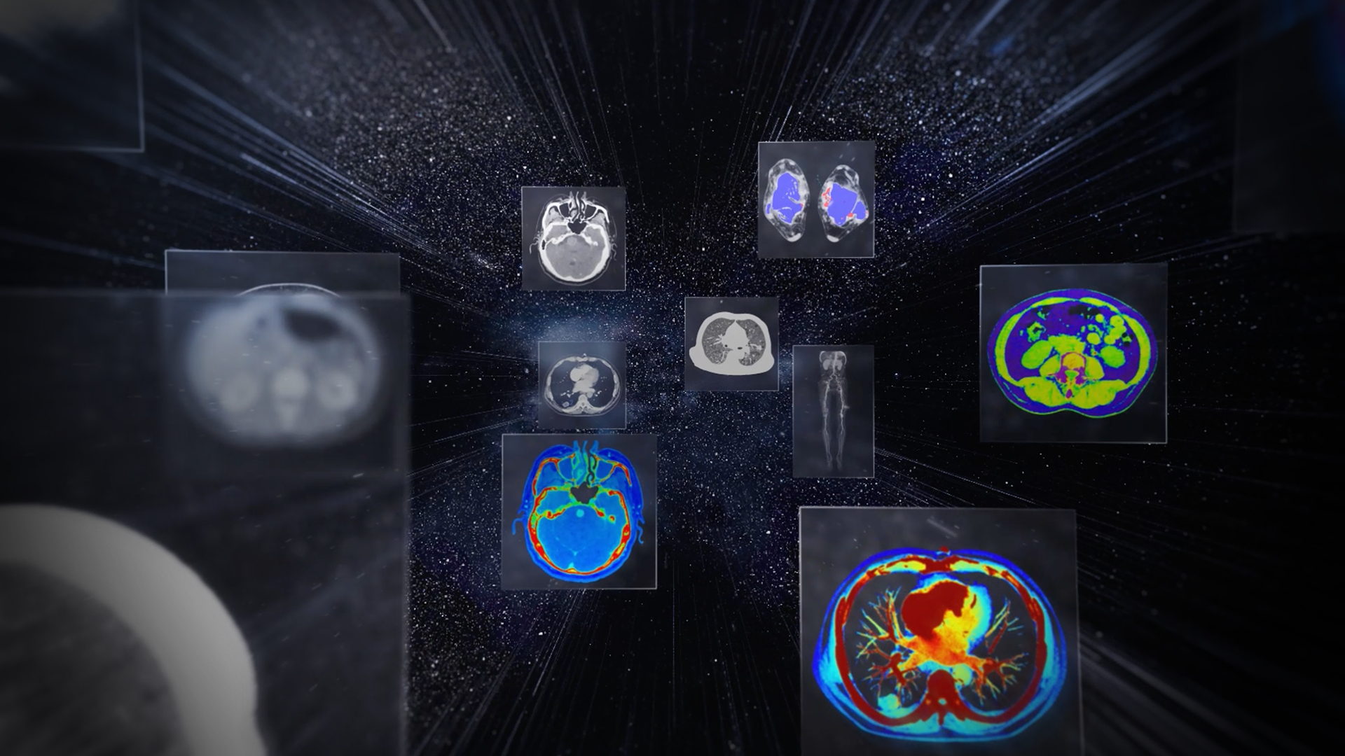
The Ultrix detector integrates the photodiode sensor array and ADC electronics through through-silicon via (TSV) technology, shortening the analog signal transmission path from centimeters to micrometers. This significantly reduces signal interference and electronic noise, enabling high-SNR, high-fidelity signal acquisition and preserving more valuable signal information for image reconstruction, particularly in low-dose imaging.
uCT SiriuX Ultrix™ features the Artificial-Intelligence Iterative Reconstruction (AIIR) , combining model-based iterative reconstruction (MBIR) with advanced deep learning denoising. By jointly modeling system optics, noise, anatomy, and physical statistics, AIIR effectively suppresses image noise and artifacts while preserving natural image texture and anatomical structures. The result is high-quality imaging at reduced radiation dose across a broad range of clinical applications.
The Ultrix detector integrates the photodiode sensor array and ADC electronics through through-silicon via (TSV) technology, shortening the analog signal transmission path from centimeters to micrometers. This significantly reduces signal interference and electronic noise, enabling high-SNR, high-fidelity signal acquisition and preserving more valuable signal information for image reconstruction, particularly in low-dose imaging.
uCT SiriuX Ultrix™ features the Artificial-Intelligence Iterative Reconstruction (AIIR) , combining model-based iterative reconstruction (MBIR) with advanced deep learning denoising. By jointly modeling system optics, noise, anatomy, and physical statistics, AIIR effectively suppresses image noise and artifacts while preserving natural image texture and anatomical structures. The result is high-quality imaging at reduced radiation dose across a broad range of clinical applications.
With 16 cm z-axis coverage and 63 ms temporal resolution, uCT SiriuX Ultrix™ enables static coronary CTA acquisition with reduced motion artifacts, supporting ECG-free coronary imaging while shortening X-ray exposure time.
uCT SiriuX Ultrix™ features CardioBoost, a deep learning reconstruction algorithm specifically optimized for cardiac imaging. Its 3D deep neural network with attention mechanism enhances critical structures such as plaques, stents, and small vessels, improving edge definition and anatomical clarity while suppressing noise and image degradation at reduced dose. CardioBoost supports high-quality cardiac imaging for coronary stenosis assessment, plaque characterization, and myocardial evaluation.
With 16 cm z-axis coverage and 63 ms temporal resolution, uCT SiriuX Ultrix™ enables static coronary CTA acquisition with reduced motion artifacts, supporting ECG-free coronary imaging while shortening X-ray exposure time.
uCT SiriuX Ultrix™ features CardioBoost, a deep learning reconstruction algorithm specifically optimized for cardiac imaging. Its 3D deep neural network with attention mechanism enhances critical structures such as plaques, stents, and small vessels, improving edge definition and anatomical clarity while suppressing noise and image degradation at reduced dose. CardioBoost supports high-quality cardiac imaging for coronary stenosis assessment, plaque characterization, and myocardial evaluation.
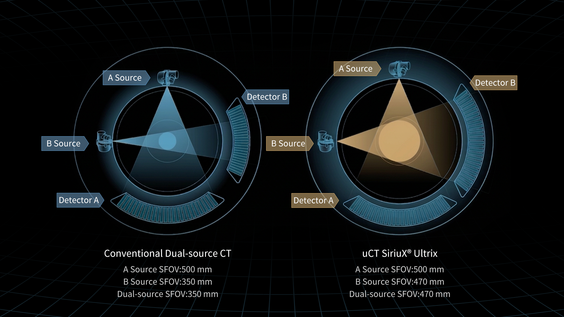
With its dual-wide coverage architecture, uCT SiriuX Ultrix™ enables high spatial resolution and advanced spectral imaging without compromise. A maximum spectral imaging field of view of 470 mm and advanced quantitative analysis support a broad range of clinical applications.
Enabled by its Twin-Ring Gantry Design, uCT SiriuX Ultrix™ expands the imaging geometry of dual-source spectral CT. Extending the detector geometry along the X–Y plane provides a maximum spectral imaging field of view of 470 mm. This enables spectral imaging across a broad anatomical range, with consistent image quality and quantitative performance.
uCT SiriuX Ultrix™ generates high- and low-energy X-rays independently from two X-ray tubes, while the PureBeam Filter minimizes spectral overlap for clear energy separation at the source. Synchronized acquisition of high- and low-energy data enables precise spectral data matching, while dual 16 cm wide-coverage detectors support 0.33 mm ultra-small-pixel spectral imaging, combining extensive anatomical coverage with exceptional structural detail. Together, these innovations establish a powerful foundation for high-fidelity spectral imaging, precise material decomposition, and reliable quantitative analysis.
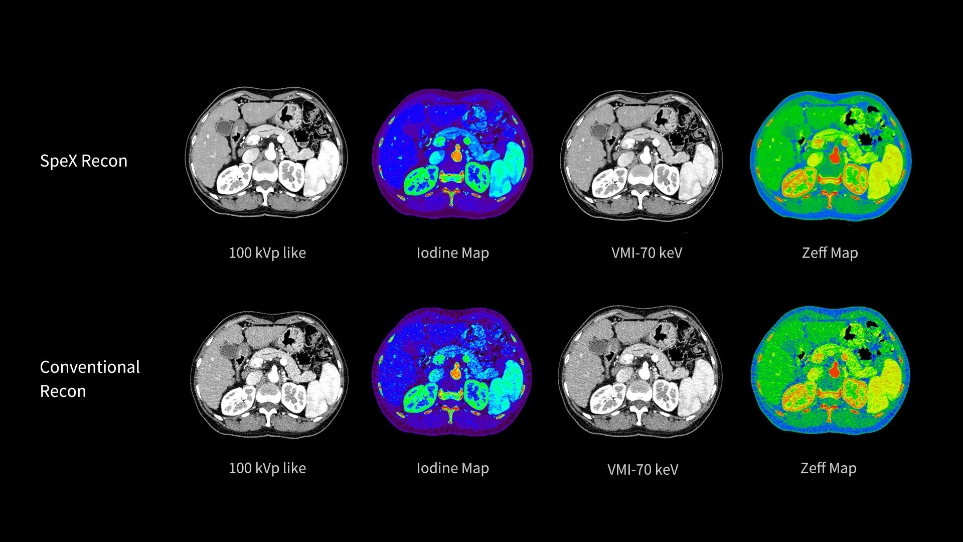
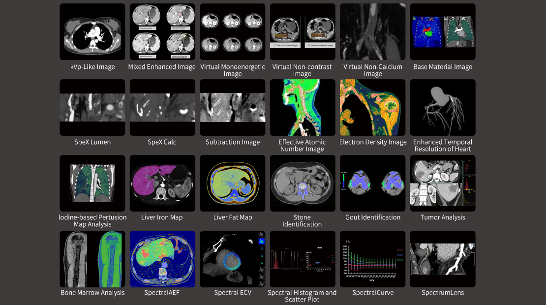
uCT SiriuX Ultrix™ Ultrix integrates SpeX Recon, an AI-powered spectral reconstruction algorithm trained on a million-level spectral image dataset. Its deep learning network directly denoises basis material images, suppressing noise while preserving fine anatomical structures and material-specific information. Based on the denoised basis material images, SpeX Recon reconstructs 15 types of spectral images, consistently carrying forward the denoising benefits to deliver optimized image quality across spectral image types, with clearer visualization and more reliable quantitative analysis.
Enabled by its Twin-Ring Gantry Design, uCT SiriuX Ultrix™ expands the imaging geometry of dual-source spectral CT. Extending the detector geometry along the X–Y plane provides a maximum spectral imaging field of view of 470 mm. This enables spectral imaging across a broad anatomical range, with consistent image quality and quantitative performance.
uCT SiriuX Ultrix™ generates high- and low-energy X-rays independently from two X-ray tubes, while the PureBeam Filter minimizes spectral overlap for clear energy separation at the source. Synchronized acquisition of high- and low-energy data enables precise spectral data matching, while dual 16 cm wide-coverage detectors support 0.33 mm ultra-small-pixel spectral imaging, combining extensive anatomical coverage with exceptional structural detail. Together, these innovations establish a powerful foundation for high-fidelity spectral imaging, precise material decomposition, and reliable quantitative analysis.
uCT SiriuX Ultrix™ Ultrix integrates SpeX Recon, an AI-powered spectral reconstruction algorithm trained on a million-level spectral image dataset. Its deep learning network directly denoises basis material images, suppressing noise while preserving fine anatomical structures and material-specific information. Based on the denoised basis material images, SpeX Recon reconstructs 15 types of spectral images, consistently carrying forward the denoising benefits to deliver optimized image quality across spectral image types, with clearer visualization and more reliable quantitative analysis.
Spectral imaging provides quantitative material-specific and energy-resolved information, with its clinical value further unlocked by an efficient spectral post-processing platform. uCT SiriuX™ offers a comprehensive suite of advanced spectral analysis tools spanning cardiology, oncology, neurology and orthopedics. With a broad range of analytical capabilities and up to 10 basis material pair image types, the platform helps transform spectral data into actionable insights for precision diagnosis and clinical research.
Dual-source Spectral Imaging
Dual-source spectral imaging delivers clear high-energy virtual monoenergetic images and efficiently suppresses metal artifacts.
PureBeam – Low-energy photon filtration
Tin Bowtie filter effectively removes low-energy photons, enhancing spectral separation in CT imaging.
DeepMAC – AI-based Metal Artifact Reduction
Powered by a deep learning network model, DeepMAC reduces artifacts from various metal implants, including hip prostheses, spinal fixation devices, orthopedic implants, dental restorations, and endovascular coils, restoring clear anatomical details.
Integrated Whole-Organ Perfusion and CTA
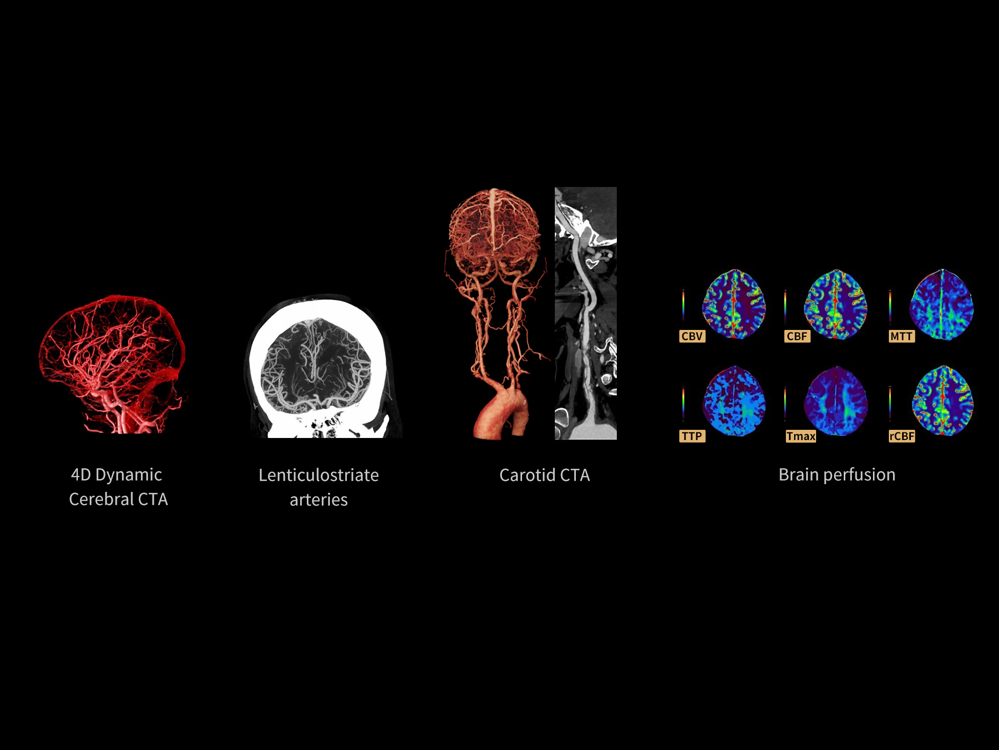
uCT SiriuX Ultrix™ eliminates the need for separate CTA and perfusion acquisitions by integrating both into a single examination. With just one contrast injection, it simultaneously delivers high-quality CTA images and quantitative whole-organ perfusion data, enabling comprehensive anatomical and functional evaluation in a streamlined workflow.
Adaptive Perfusion Scan
Real-time monitoring automatically triggers CTA acquisition and completes CTP at the optimal time point, enabling high-quality CTA and quantitative perfusion analysis in a single examination while optimizing radiation dose.
Integrated Perfusion and Spectral Imaging
Flexible spectral acquisition throughout the perfusion study enables full-phase spectral perfusion imaging or targeted arterial-phase acquisition, simultaneously delivering high-quality CTA images and quantitative iodine kinetics.
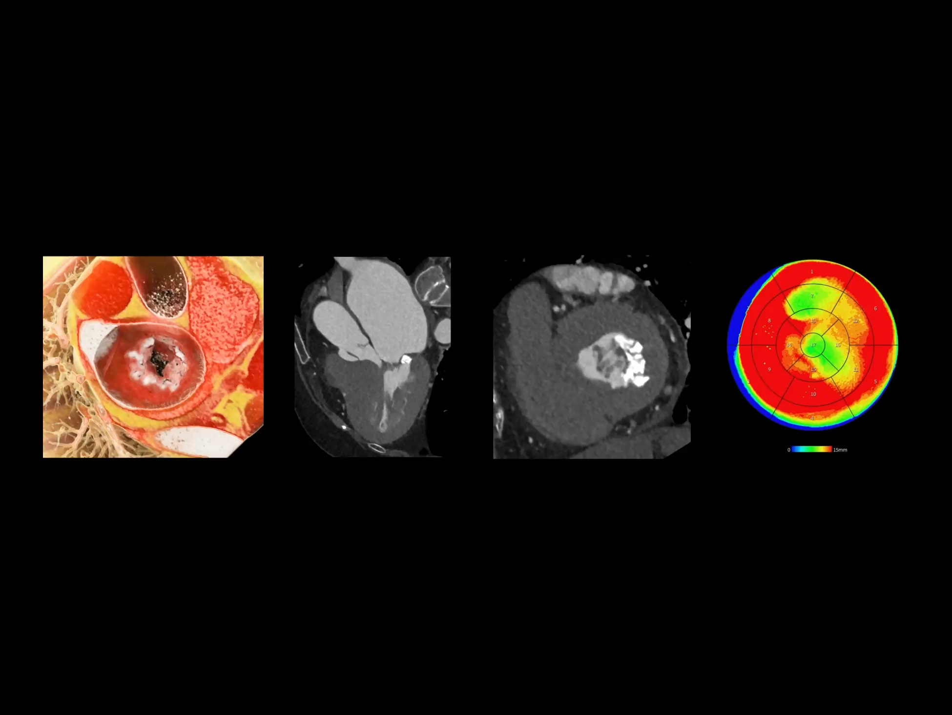
Cardiac Imaging
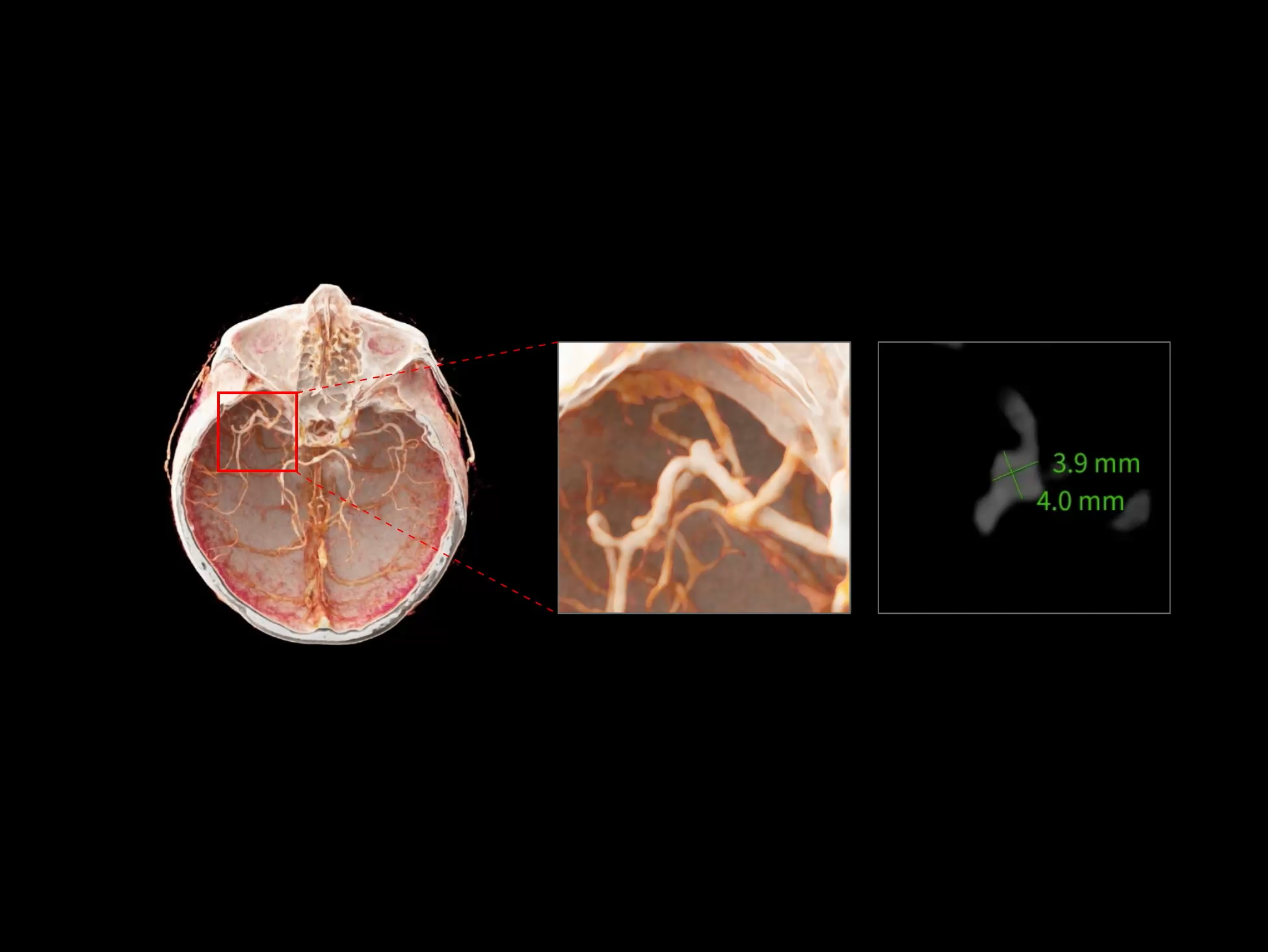
Head and Neck Imaging
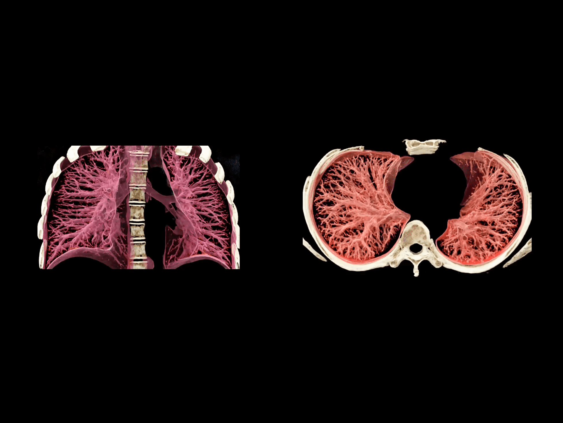
Chest Imaging
Abdominal Imaging
Musculoskeletal Imaging
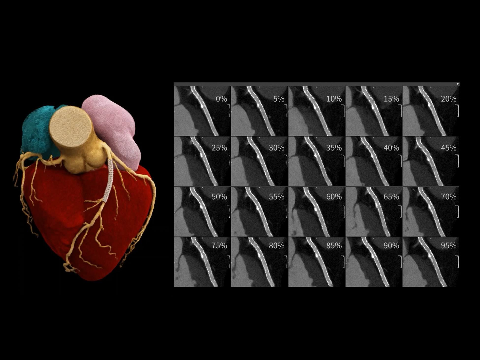
0.33 mm x 0.33 mm, 1024 x 1024
100 kV, 97 mAs, 14.18 mGy, Effective Dose: 2.79 mSv
Detailed Dynamic Visualization of Coronary Stent Morphology
0.33 mm x 0.33 mm, 1024 x 1024 matrix
100 kV, 127 mAs, 19.69 mGy, Effective Dose: 3.87 mSv
Dynamic Assessment of Annuloplasty with Artificial Chordae Implantation
0.33 mm x 0.33 mm, 1024 x 1024
CCTA: 100 kV, 96 mAs, 14.62 mGy, Effective Dose: 2.35 mSv
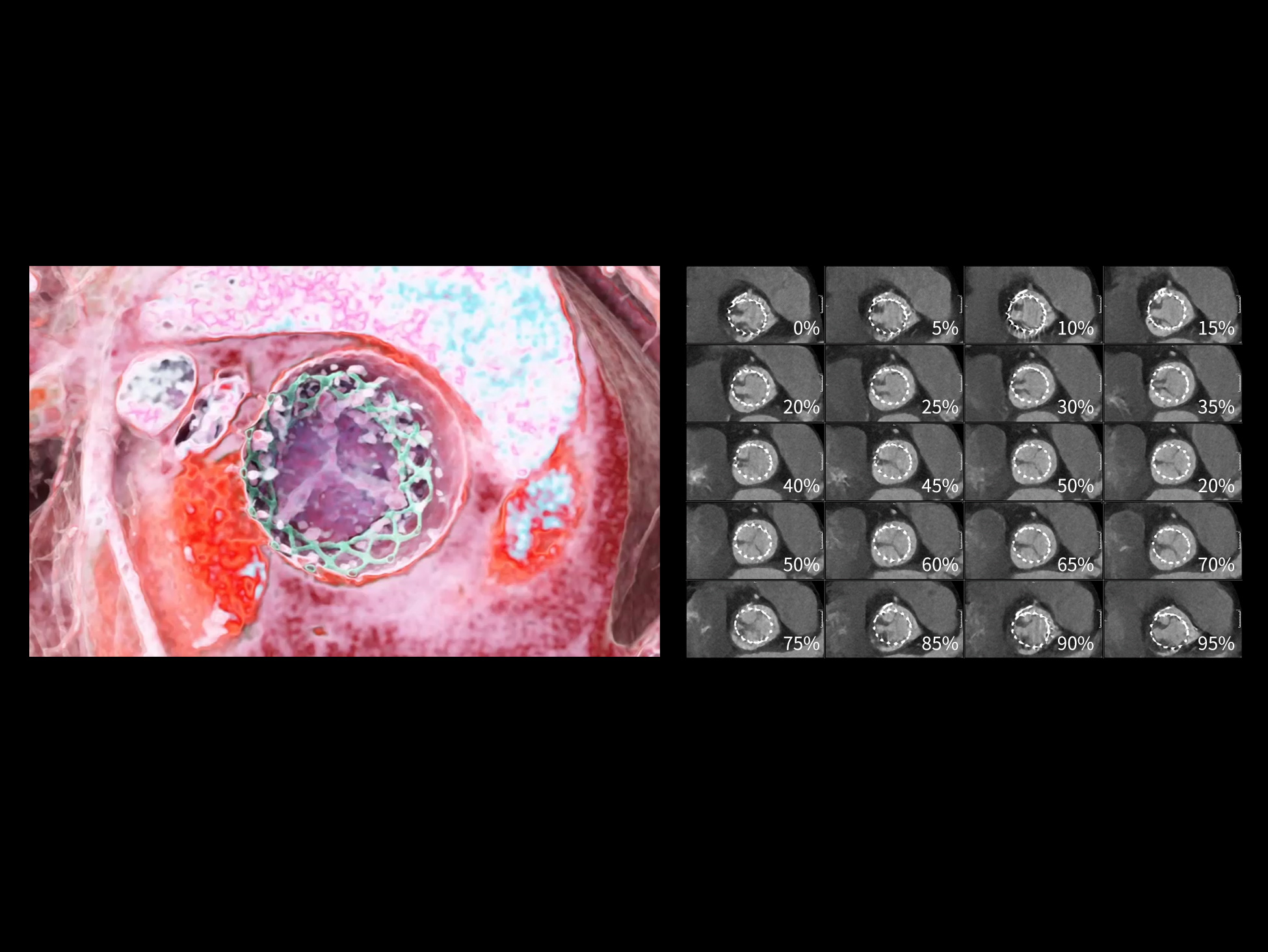
Full Cardiac Cycle Evaluation of the TAVR Follow-up Study
100 kV, 132 mAs, 8.37 mGy, Effective Dose: 1.87 mSv
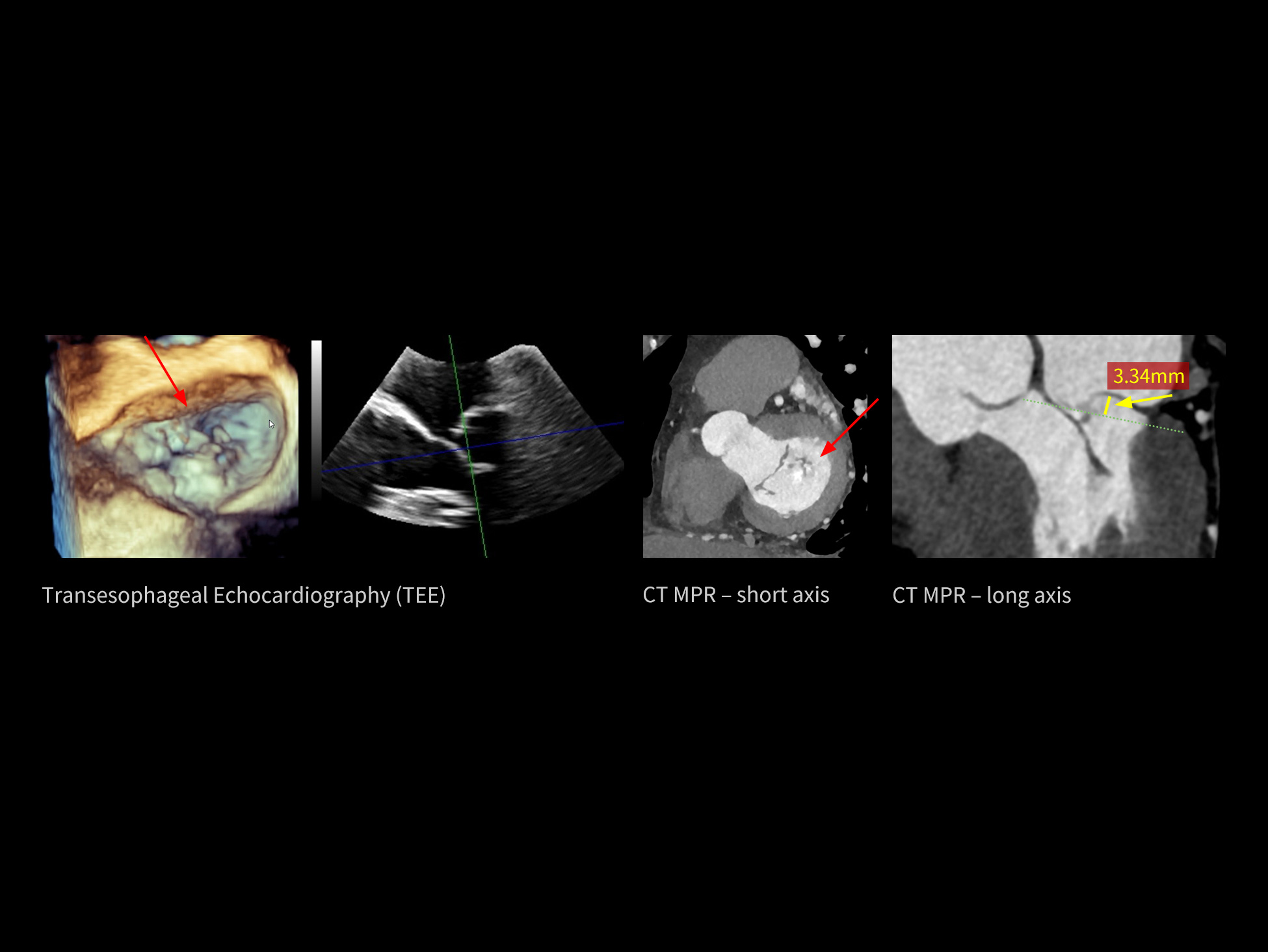
Dynamic Mitral Valve Prolapse (MVP) Evaluation
100 kV, 95 mAs, 14.19 mGy, Effective Dose: 3.12 mSv
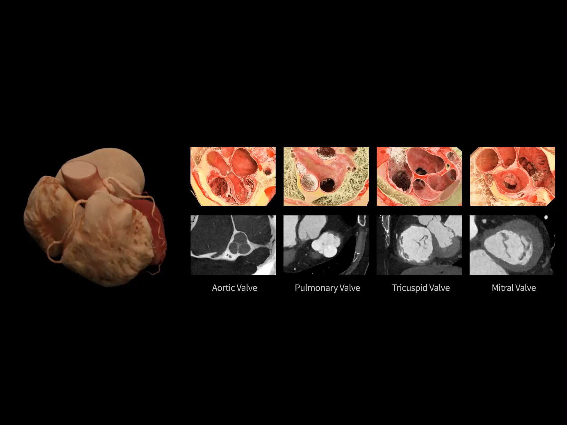
Simultaneous High-Definition Visualization of All Four Valves and Chordae Tendineae
100 kV, 106 mAs, 16.61 mGy, Effective Dose: 3.27 mSv
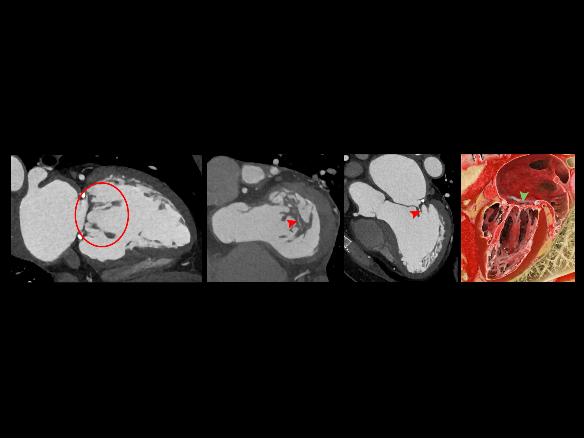
Assessment of Hypertrophic Cardiomyopathy with Severe Mitral Annular Calcification and Dynamic Stenosis
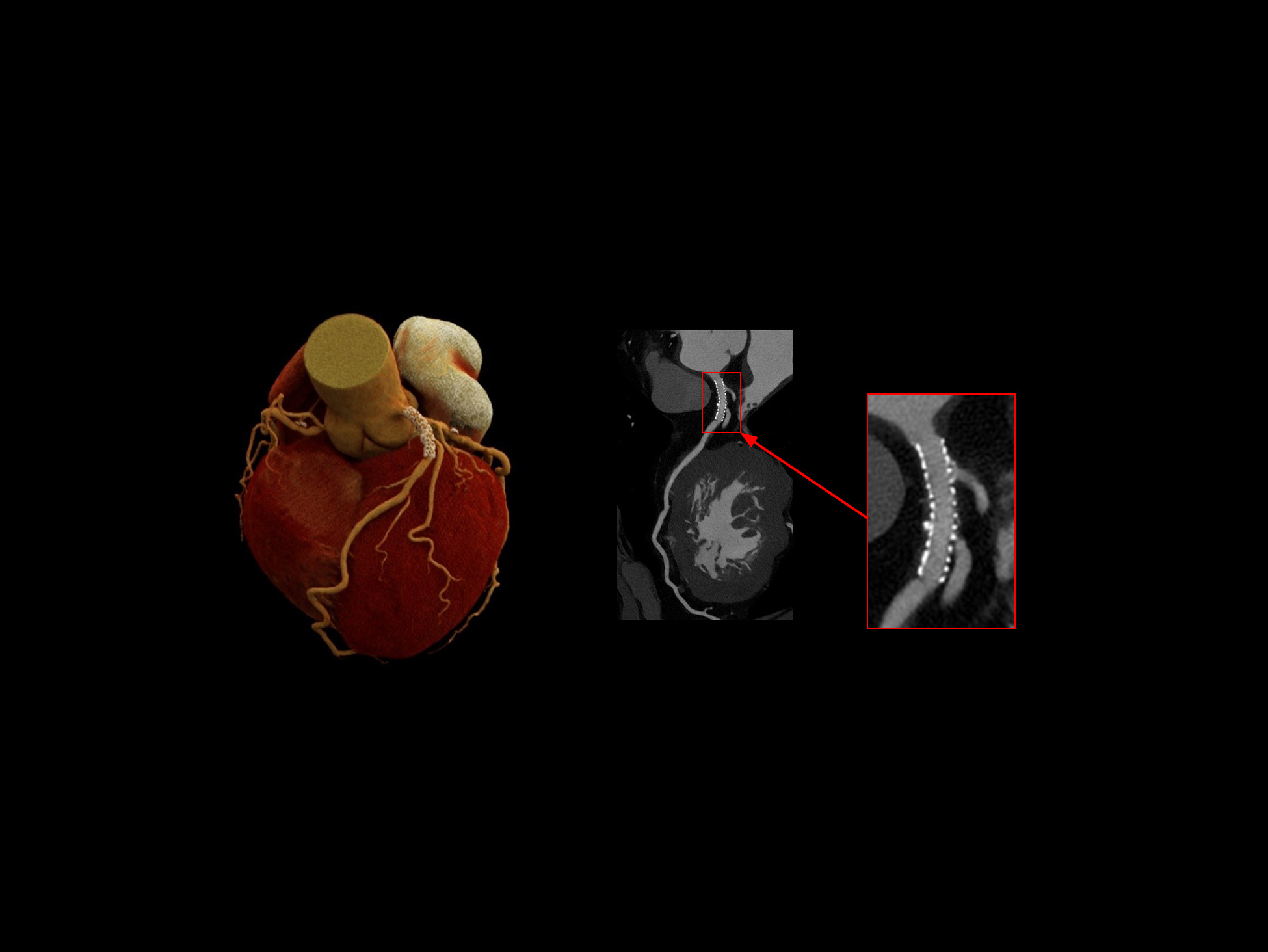
100 kV, 125 mAs, 4.42 mGy, Effective Dose: 0.87 mSv
Stent architecture and luminal patency clearly visualized
100 kV, 100 mAs, 13.82 mGy, Effective Dose: 0.47mSv
Full Cardiac Cycle Evaluation of the Brain Aneurysm
CTP: 80 kV, 119 mAs, 38.7 mGy, Effective Dose: 3.2 mSv
CTA: 80 kV, 298 mAs, 5.18 mGy, Effective Dose: 0.49 mSv
A Comprehensive Workflow for Anatomical and Hemodynamic Cerebrovascular Assessment
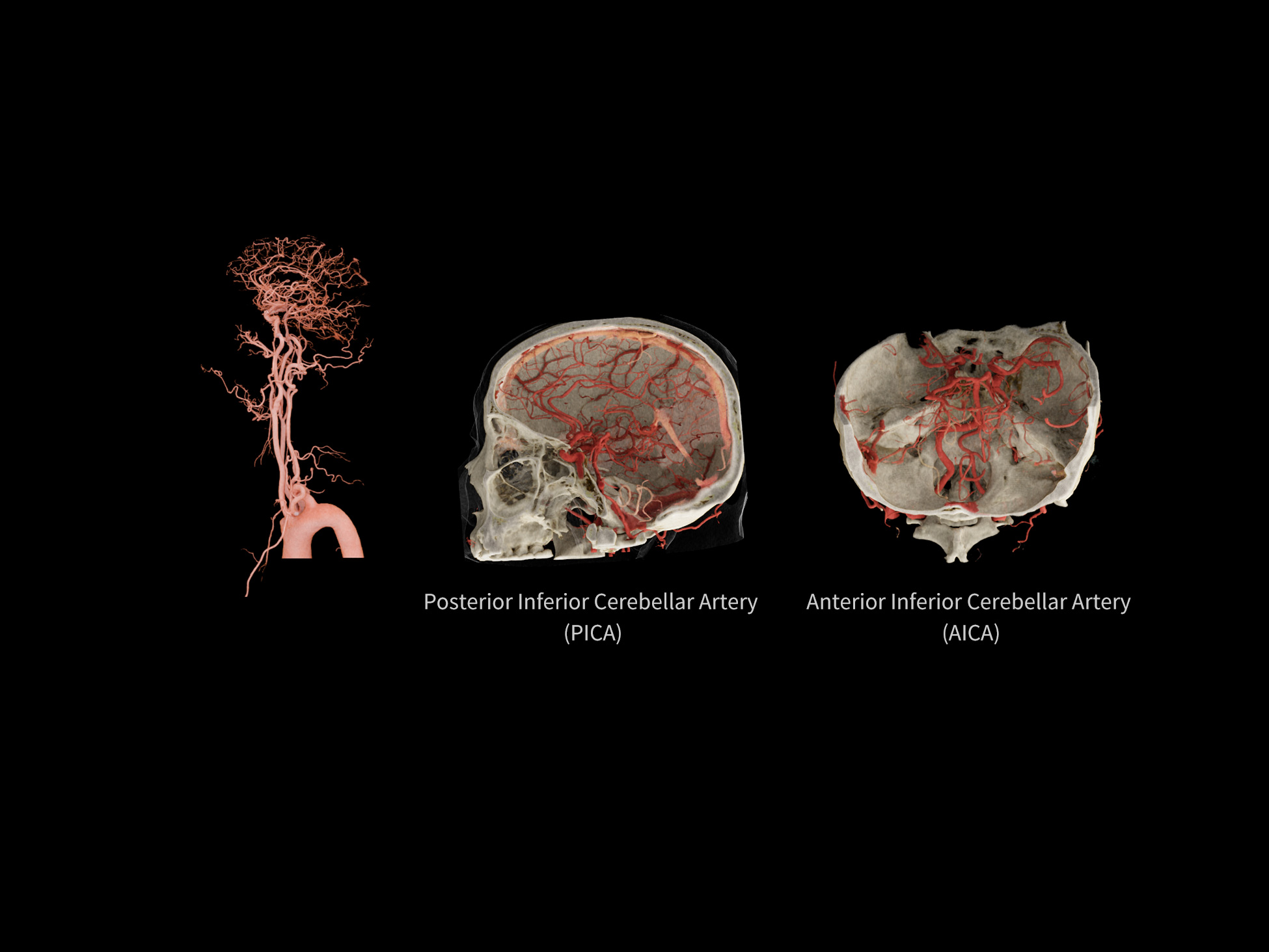
0.33 mm x 0.33 mm, 1024 x 1024
100 kV, 213 mAs, 7.70 mGy, Effective Dose: 1.2 mSv
Enabling Accurate Evaluation of Cerebral Microvascular Pathology
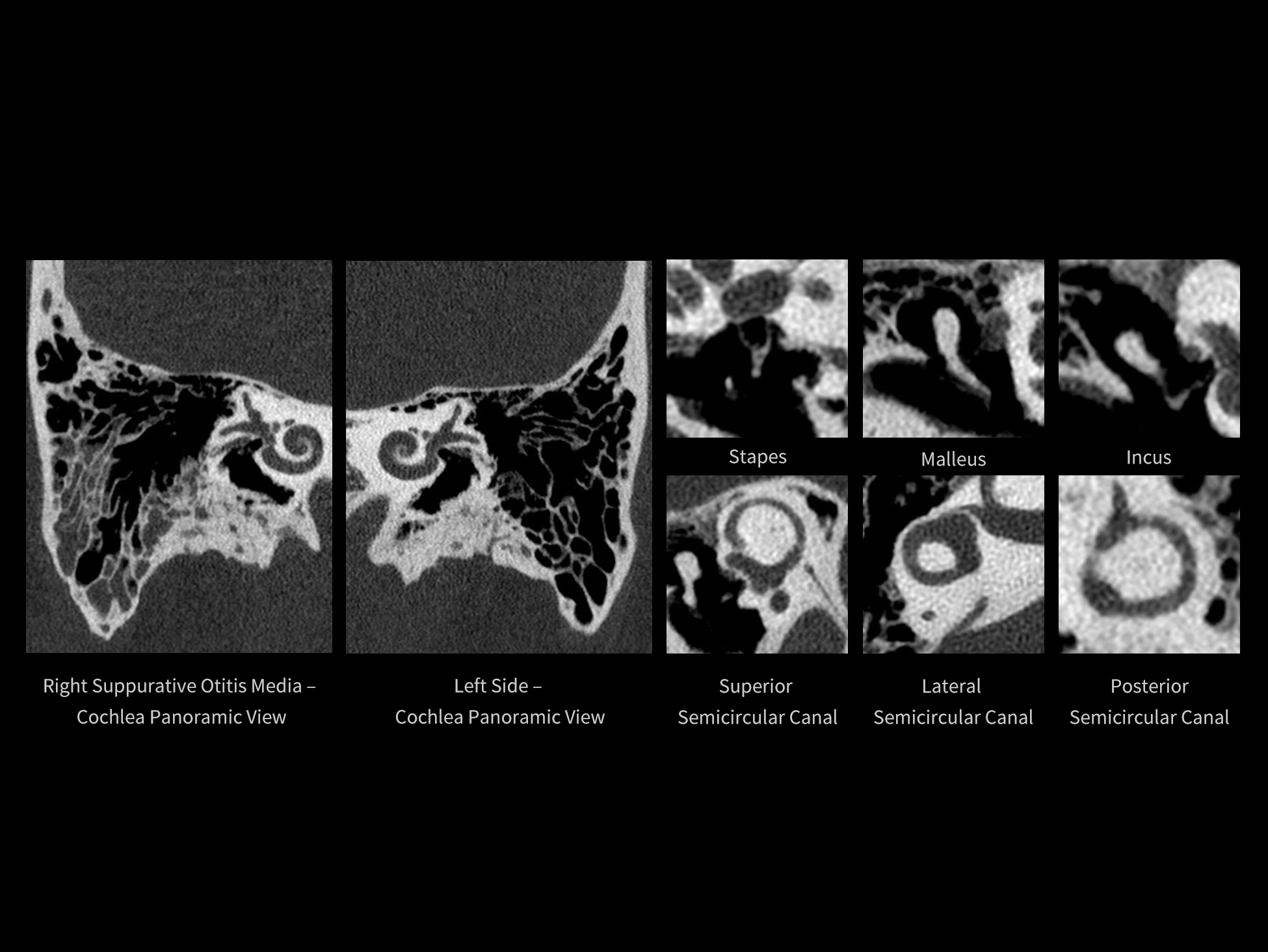
0.17 mm x 0.08 mm, 2048 x 2048
140 kV, 149 mAs, 32.88 mGy, Effective Dose: 0.84 mSv
High-Fidelity Visualization of Cochleovestibular Microstructures
0.33 mm x 0.33 mm, 1024 x 1024
120 kV, 10 mAs, 5.79 mGy, 1.30 mSv
Tracking Respiratory Motion, Lung Volume, and Fine Bronchial-Vascular Structures
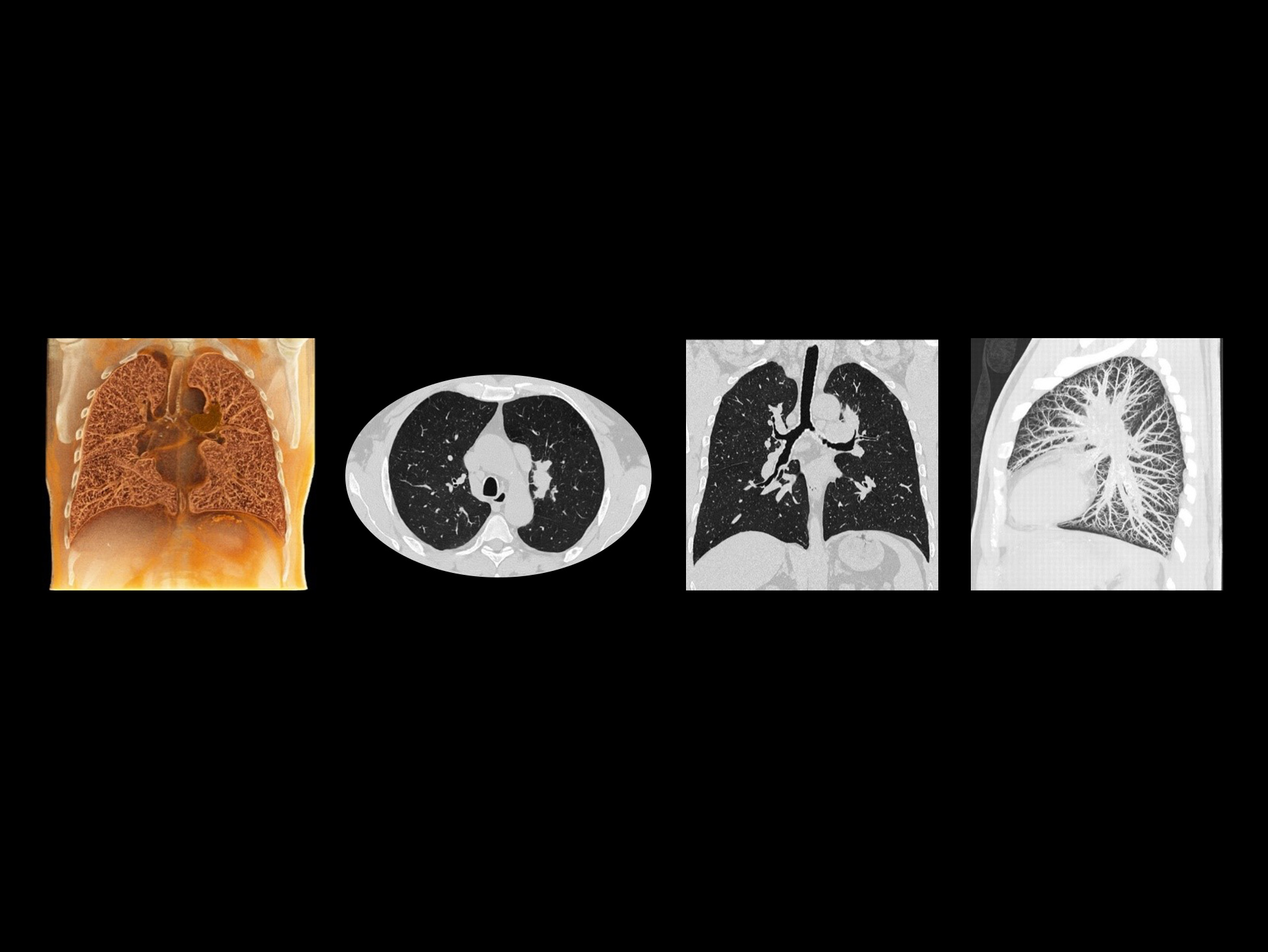
120 kV, 50 mAs, 3.00 mGy, Effective Dose: 1.9 mSv
Clearly Visualizing Fine Bronchial and Vascular Structures
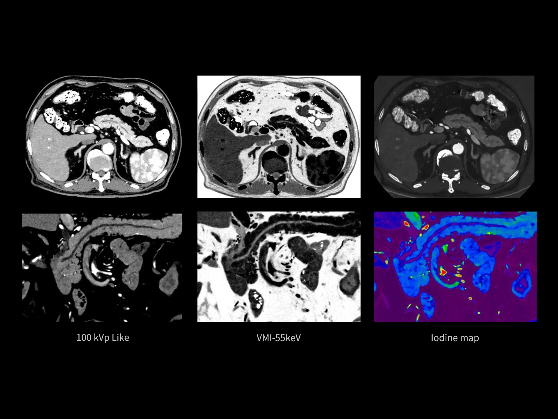
0.33 mm x 0.33 mm, 768× 768
80 kV/140 kV Tin,6.13 mGy, Effective Dose: 4.14 mSv
Delineating the Full Extent of Pancreatic Ductal Anatomy
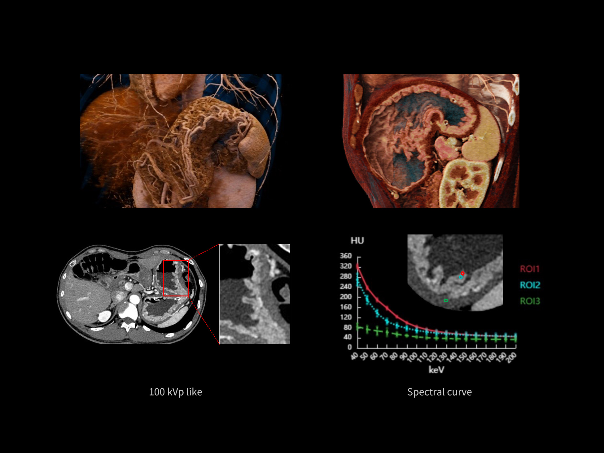
0.33 mm x 0.17 mm, 512 x 512
80 kV/140 kV Tin,6.07 mGy, Effective Dose: 3.28 mSv
Revealing Fine Gastric Wall Layers and Small Vascular Branches
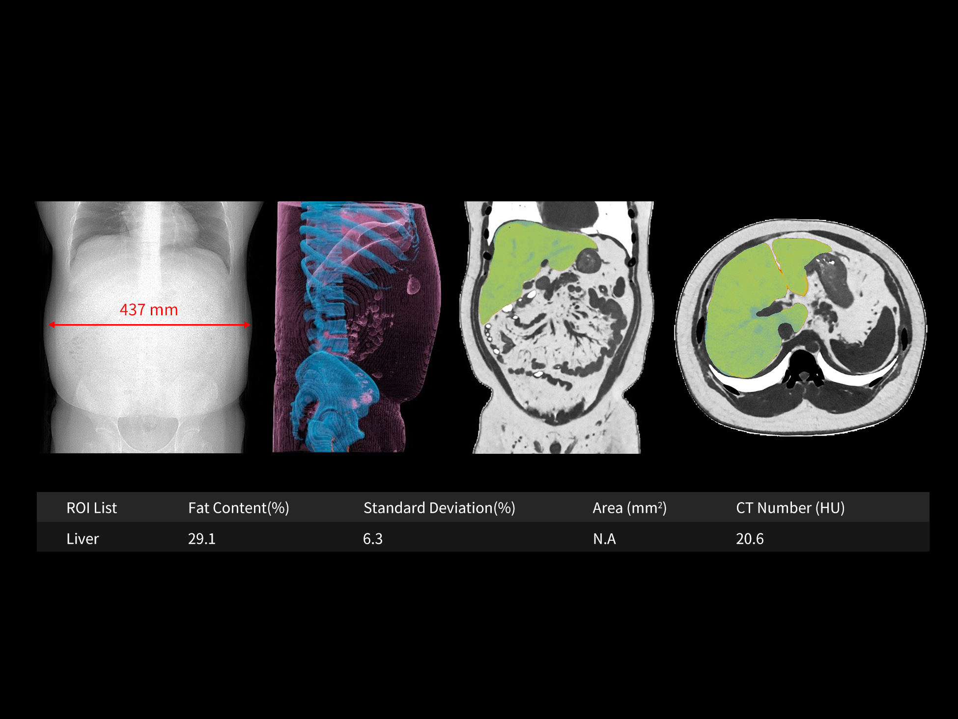
80 kV/140 kV Tin,14.18 mGy, Effective Dose: 11.60 mSv
Full Abdominal Coverage for High-Definition Imaging and Hepatic Fat Quantification
120 kV, 23 mAs, 15.35 mGy, Effective Dose: 3.69 mSv
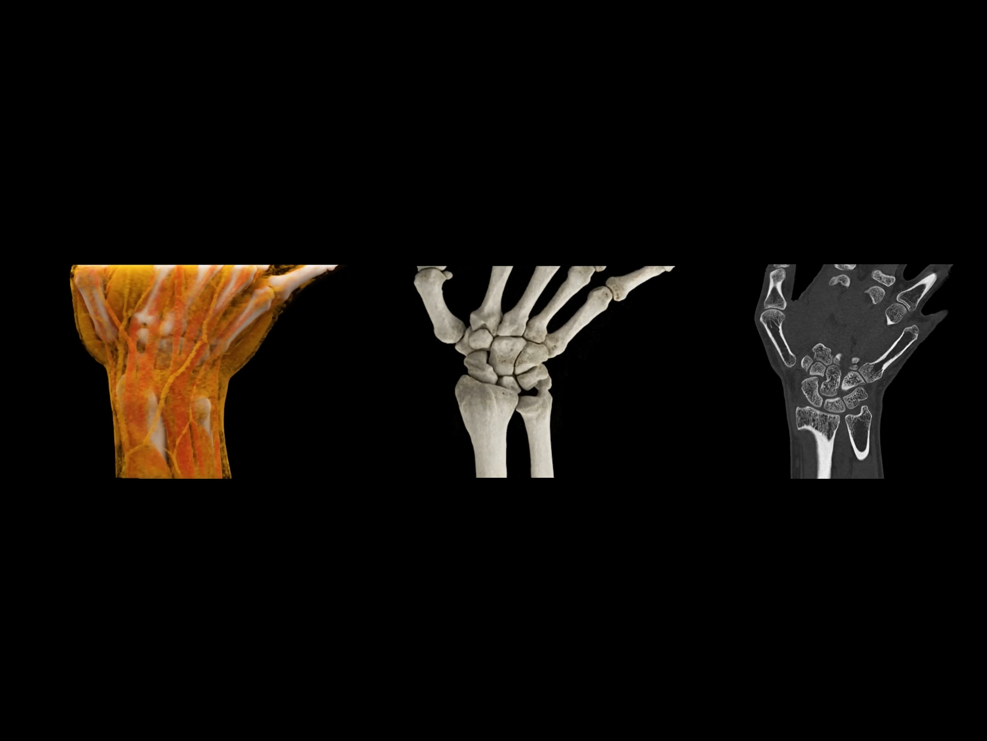
Dynamic Assessment of Wrist Stability
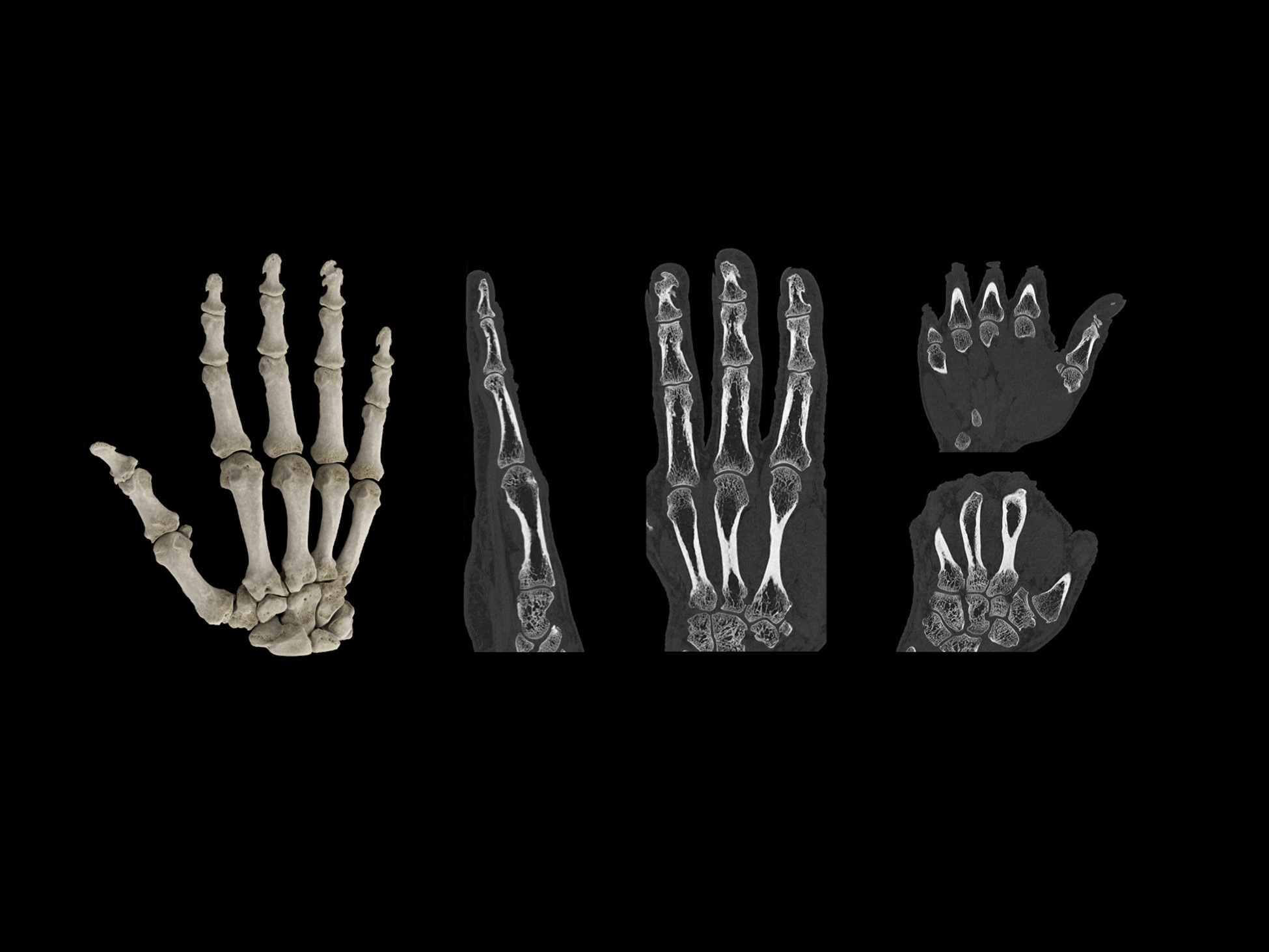
0.17mm X 0.17mm, 1024 x 1024 matrix
120 kV, 60 mAs, 4.85 mGy, Effective dose: 1.6 mSv
Clear Visualization of Trabecular and Cortical Microarchitecture
|
Gantry bore size |
82 cm |
|
Detectors |
2x16 cm Ultrix™ Detectors |
|
Number of slices per rotation |
2x960 Slices |
|
Pixel size @ Iso-center |
0.33 mm |
|
Minimum reconstruction slice thickness |
0.17 mm |
|
X-ray tubes |
2x 34 MHU Liquid Metal Bearing X-ray Tubes |
|
Rotation time |
Up to 0.239 sec/rotation |
|
Temporal resolution |
Up to 8 ms with CardioCapture |
|
Table load |
Up to 325 kg |
|
kV settings |
60/70/80/100/120/140 kV |
|
Generator power |
2x100 kW |
Disclaimer:
The product may not be commercially available in all countries. Please check with your local United Imaging Healthcare representative for availability.
This product is intended for use by trained medical professionals only. Patients should follow the guidance and instructions provided by healthcare practitioners during operation.
The supported language of the instructions for use is English. Qualified users may access the instruction for use in the appropriate language via the provided link: https://uicloud.com/uclass/docs/en-us/home
Please contact your local United Imaging Healthcare organization for further details.