

Vertices Unite, Innovations Ignite

CT continues to evolve, moving beyond static anatomy to reveal the dynamics of human physiology. With its dual-wide coverage architecture, uCT SiriuX™ combines ultra-fast temporal resolution, extensive volumetric coverage, and advanced spectral imaging. It enables clinicians to see more, understand more, and capture the dynamics of life.

Pioneering Unprecedented Dynamics


Built on a dual-wide coverage architecture, uCT SiriuX™ delivers both ultra-high temporal resolution and extensive volumetric coverage, unlocking new possibilities in CT performance. It precisely captures the full spectrum of physiological motion, offering a new perspective on dynamic imaging.

Dual-wide Coverage Architecture

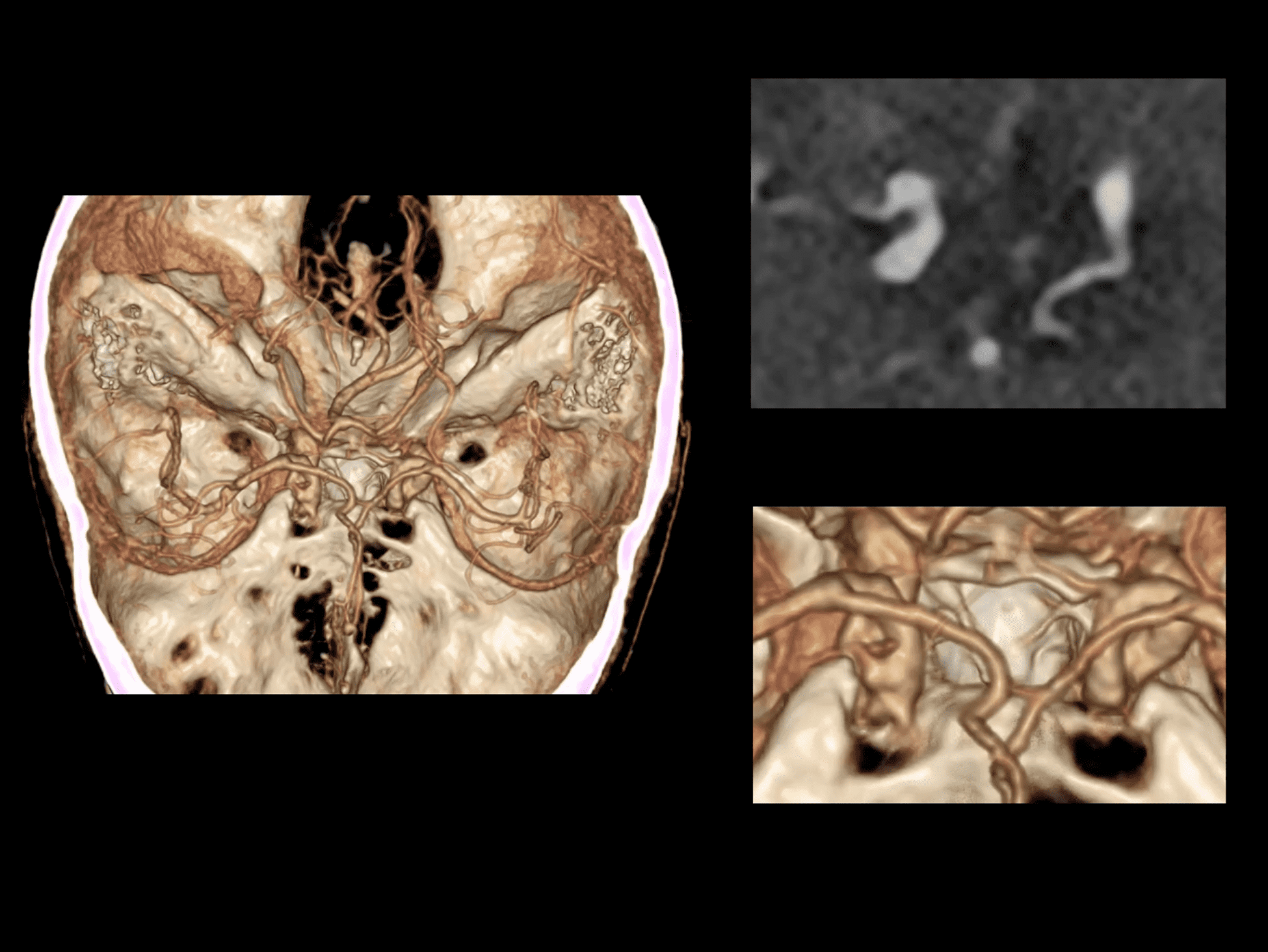
Powered by dual detectors with 16 cm Z-axis coverage, uCT SiriuX™ enables comprehensive dynamic imaging from brain to heart and beyond. Combined with ultra-high temporal resolution, it delivers sharper, faster, and more precise visualization of physiological motion.

Built on a dual-source CT architecture, uCT SiriuX™ achieves enhanced physical temporal resolution for capturing rapid physiological changes with exceptional clarity. Liquid metal bearing technology ensures smooth and stable rotation, while the 34 MHU high-capacity anode supports sustained performance under demanding clinical workloads.

Twin-ring gantry design redefines CT architecture. The imaging ring integrates dual X-ray tubes and dual 16 cm wide-coverage detectors, while the carrier ring integrates power and thermal management for a compact, efficient design. The separable Twin-rings simplify installation and maintenance. With micron-level precision and high-speed data synchronization, uCT SiriuX™ delivers reliable, high-precision imaging for every scan.

2x16 cm Wide-coverage Detectors

Powered by dual detectors with 16 cm Z-axis coverage, uCT SiriuX™ enables comprehensive dynamic imaging from brain to heart and beyond. Combined with ultra-high temporal resolution, it delivers sharper, faster, and more precise visualization of physiological motion.

2x Liquid Metal Bearing X-ray Tubes

Built on a dual-source CT architecture, uCT SiriuX™ achieves enhanced physical temporal resolution for capturing rapid physiological changes with exceptional clarity. Liquid metal bearing technology ensures smooth and stable rotation, while the 34 MHU high-capacity anode supports sustained performance under demanding clinical workloads.

Twin-ring Gantry Design

Twin-ring gantry design redefines CT architecture. The imaging ring integrates dual X-ray tubes and dual 16 cm wide-coverage detectors, while the carrier ring integrates power and thermal management for a compact, efficient design. The separable Twin-rings simplify installation and maintenance. With micron-level precision and high-speed data synchronization, uCT SiriuX™ delivers reliable, high-precision imaging for every scan.

Enabling Whole-organ Synchronized Temporal Resolution

Building on a dual-source architecture, uCT SiriuX™ adopts an advanced gantry design to overcome the challenges of increased rotational inertia and high centrifugal forces, enabling fast rotation and an ultra-high physical temporal resolution of 63 ms.

0.239 s/360°
Rotation Speed

63 ms
Native Temporal Resolution

With 2 × 16 cm wide Z-axis coverage, uCT SiriuX™ combines ultra-fast temporal resolution with whole-organ coverage to capture physiological motion in its entirety. From cardiac motion to joint movement, complete anatomical regions can be imaged simultaneously with exceptional clarity, minimizing temporal mismatch and advancing CT from partial coverage to whole-organ synchronized imaging.

Ultra-fast Temporal Resolution

Building on a dual-source architecture, uCT SiriuX™ adopts an advanced gantry design to overcome the challenges of increased rotational inertia and high centrifugal forces, enabling fast rotation and an ultra-high physical temporal resolution of 63 ms.

0.239 s/360°
Rotation Speed

63 ms
Native Temporal Resolution

Whole-Organ Synchronization

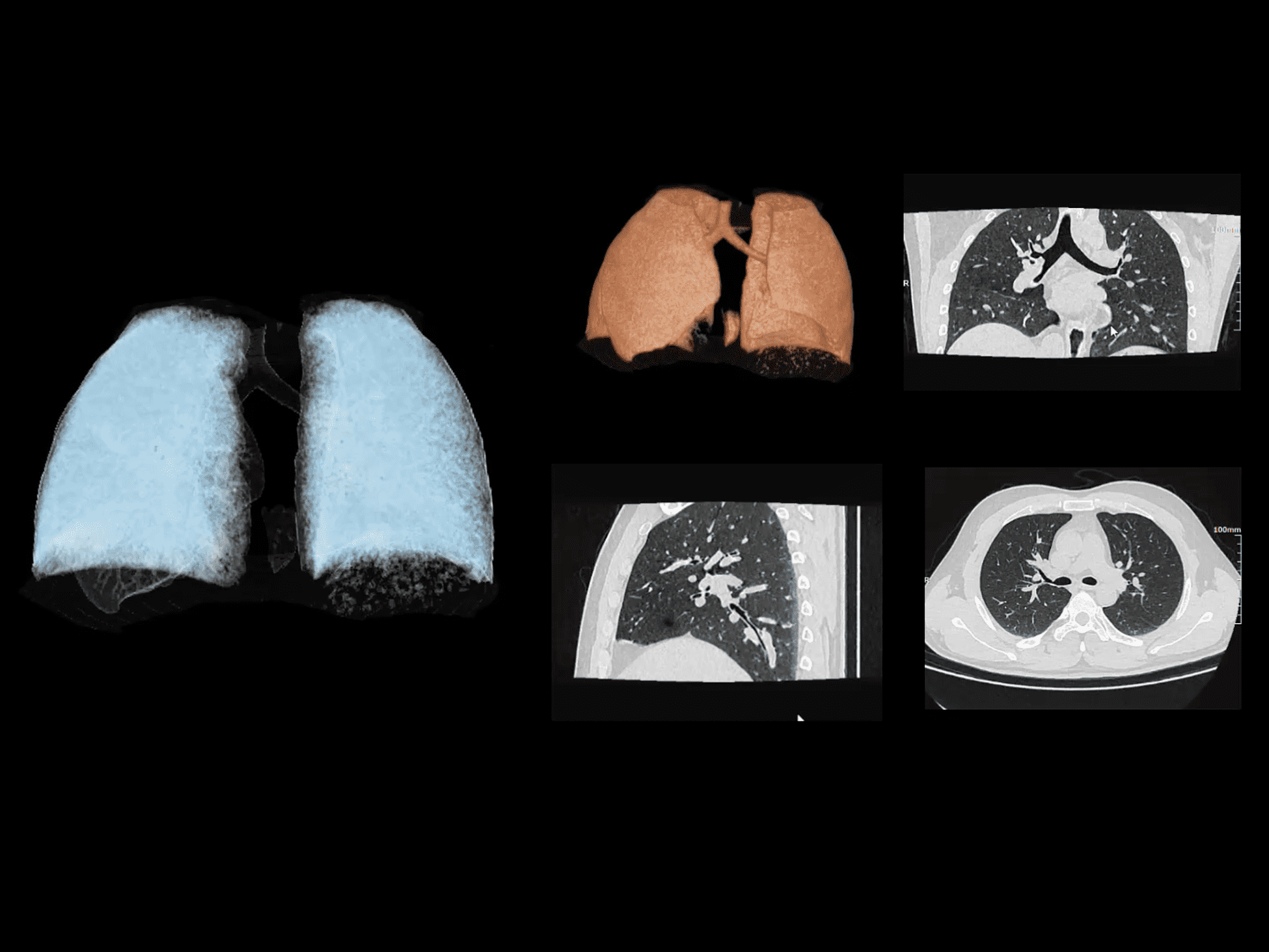
With 2 × 16 cm wide Z-axis coverage, uCT SiriuX™ combines ultra-fast temporal resolution with whole-organ coverage to capture physiological motion in its entirety. From cardiac motion to joint movement, complete anatomical regions can be imaged simultaneously with exceptional clarity, minimizing temporal mismatch and advancing CT from partial coverage to whole-organ synchronized imaging.

Unprecedented Whole-body Dynamic Imaging

Revolutionizing Whole-heart Imaging


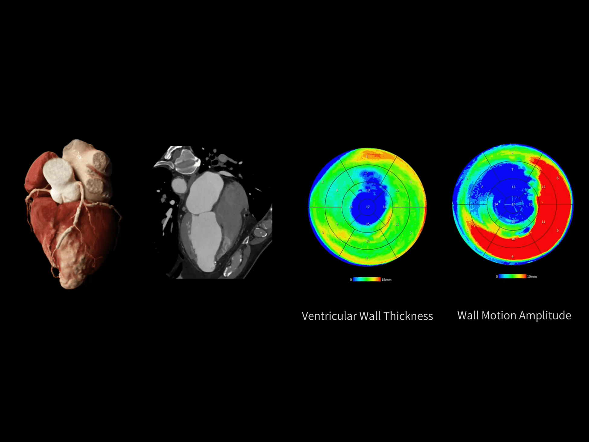
uCT SiriuX™ pushes the boundaries of cardiac temporal resolution, precisely capturing dynamic changes throughout the entire cardiac cycle. From static anatomical structures to dynamic functional performance, every detail is revealed with millisecond-level temporal precision.

Pushing the Boundaries of Whole-heart Temporal Resolution

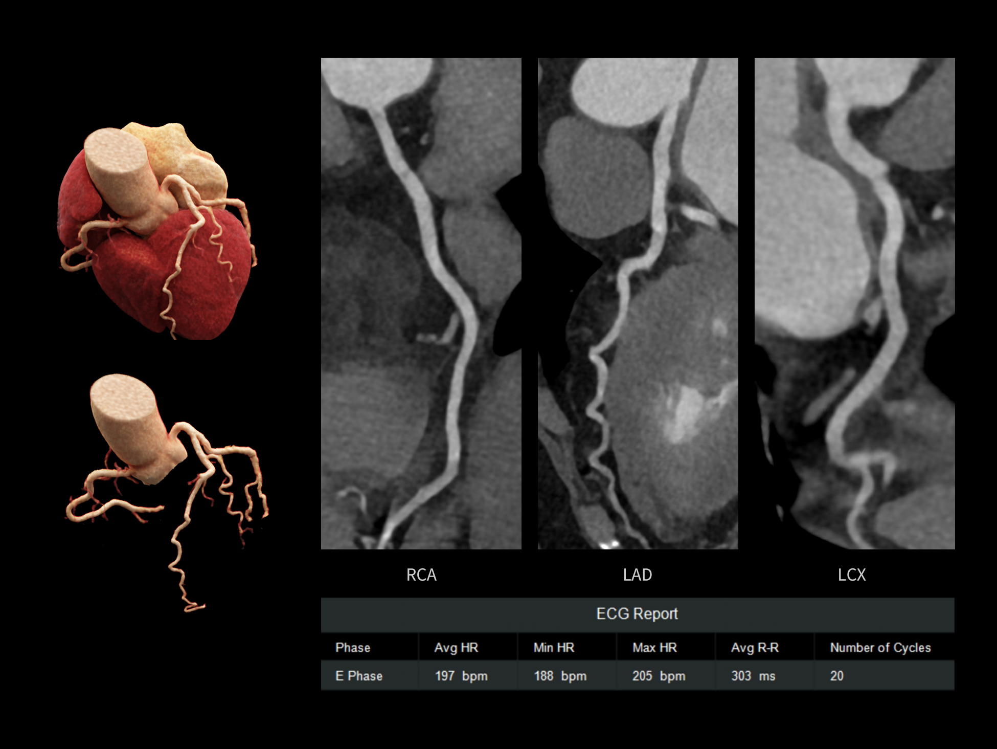
CardioCapture 2.0 achieves an effective temporal resolution of 8 ms through AI-powered cardiac motion correction. By precisely segmenting cardiac structures, tracking motion trajectories, and generating multi-phase motion fields, it enables synchronized motion correction of the coronary arteries, valves, myocardium, and ventricles for clearer visualization of cardiac dynamics throughout the entire cardiac cycle.

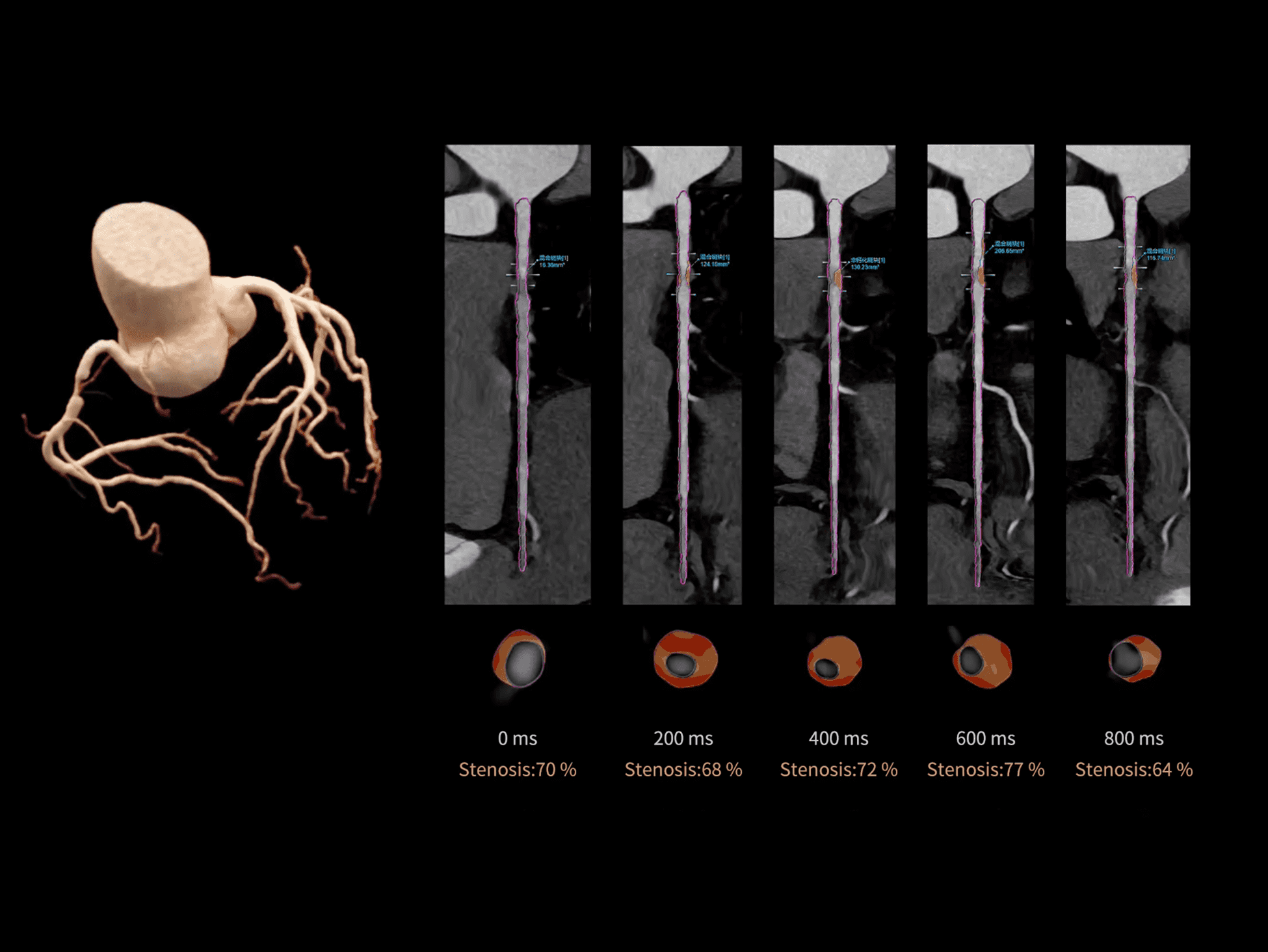
CardioCapture 2.0 reconstructs multi-phase images before and after the target phase, using deep learning networks to accurately extract coronary centerlines and establish inter-phase motion models. This enables precise motion correction of segmented coronary images for clearer coronary visualization.

CardioCapture 2.0 provides synchronized motion correction for the myocardium, valves, and ventricles. Powered by deep learning-based cardiac segmentation, it extracts comprehensive cardiac information, effectively suppresses motion artifacts, and significantly enhances structural clarity and diagnostic confidence.

8 ms Whole-heart Temporal Resolution

CardioCapture 2.0 achieves an effective temporal resolution of 8 ms through AI-powered cardiac motion correction. By precisely segmenting cardiac structures, tracking motion trajectories, and generating multi-phase motion fields, it enables synchronized motion correction of the coronary arteries, valves, myocardium, and ventricles for clearer visualization of cardiac dynamics throughout the entire cardiac cycle.

Coronary Artery Motion Correction

CardioCapture 2.0 reconstructs multi-phase images before and after the target phase, using deep learning networks to accurately extract coronary centerlines and establish inter-phase motion models. This enables precise motion correction of segmented coronary images for clearer coronary visualization.

Whole-heart Motion Correction

CardioCapture 2.0 provides synchronized motion correction for the myocardium, valves, and ventricles. Powered by deep learning-based cardiac segmentation, it extracts comprehensive cardiac information, effectively suppresses motion artifacts, and significantly enhances structural clarity and diagnostic confidence.

Expanding the Possibilities of Cardiac Imaging

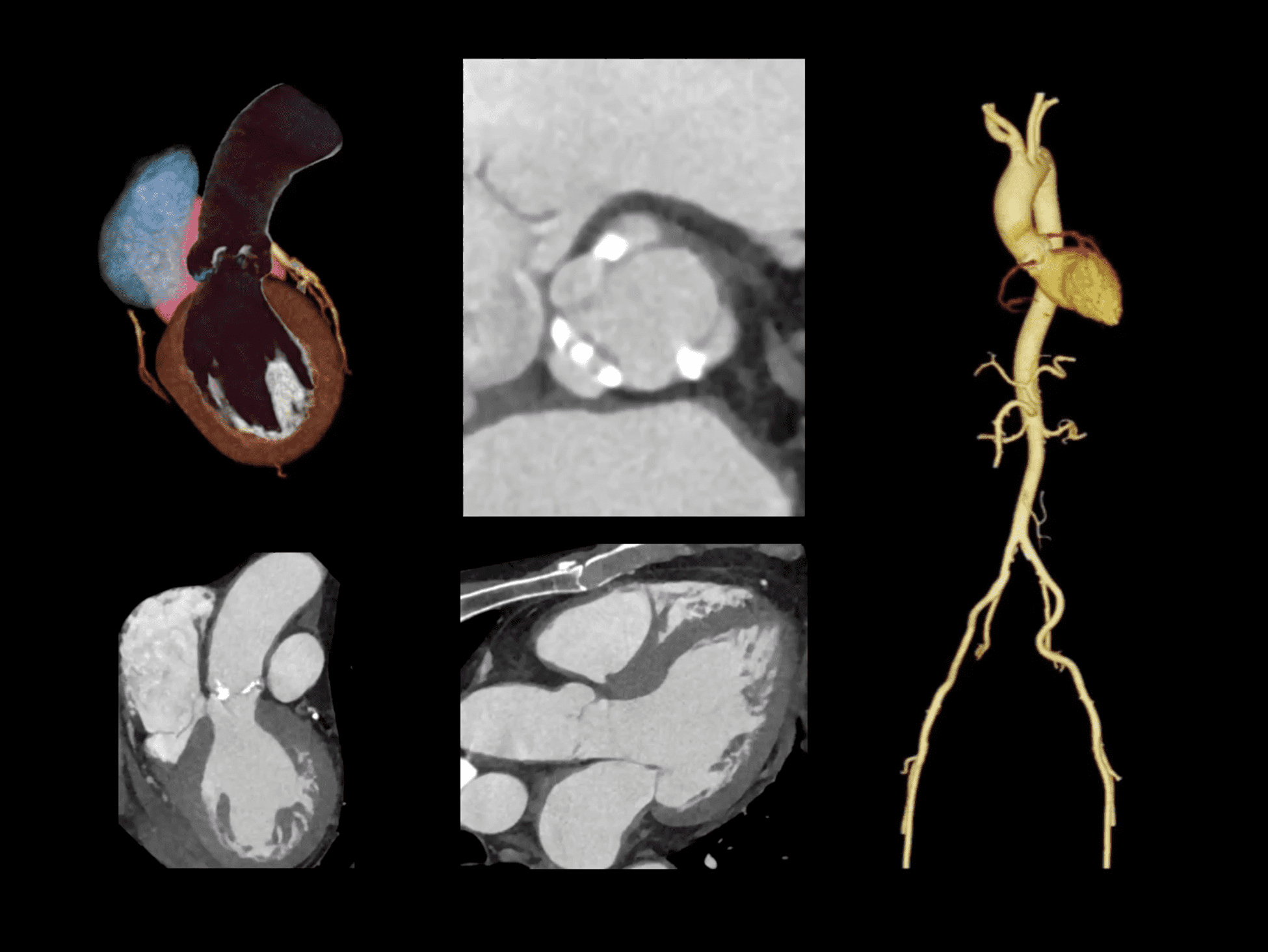
With 8 ms ultra-high temporal resolution and 16 cm wide coverage, uCT SiriuX™ captures the complete anatomy and dynamic function of the beating heart beyond the limits of conventional cardiac imaging. Combined with ECG-free scanning and intelligent workflows, it reduces the impact of heart rate, patient cooperation, and operational complexity, delivering a more flexible and confident cardiac imaging experience.

4D Whole-heart Dynamic Imaging

Captures cardiac motion throughout the entire cardiac cycle, providing high-definition visualization of dynamic structural and functional changes.

Whole-heart Full-phase Imaging

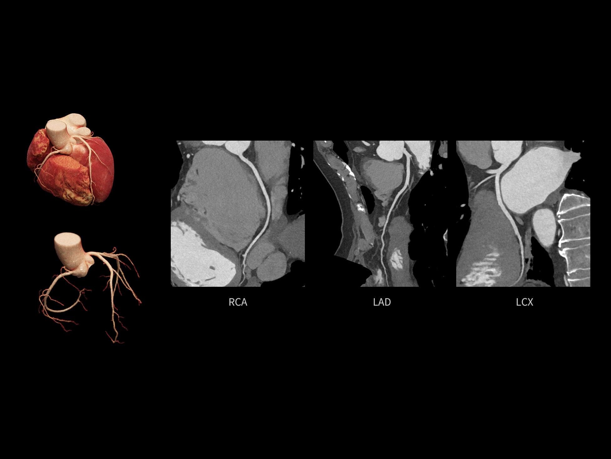
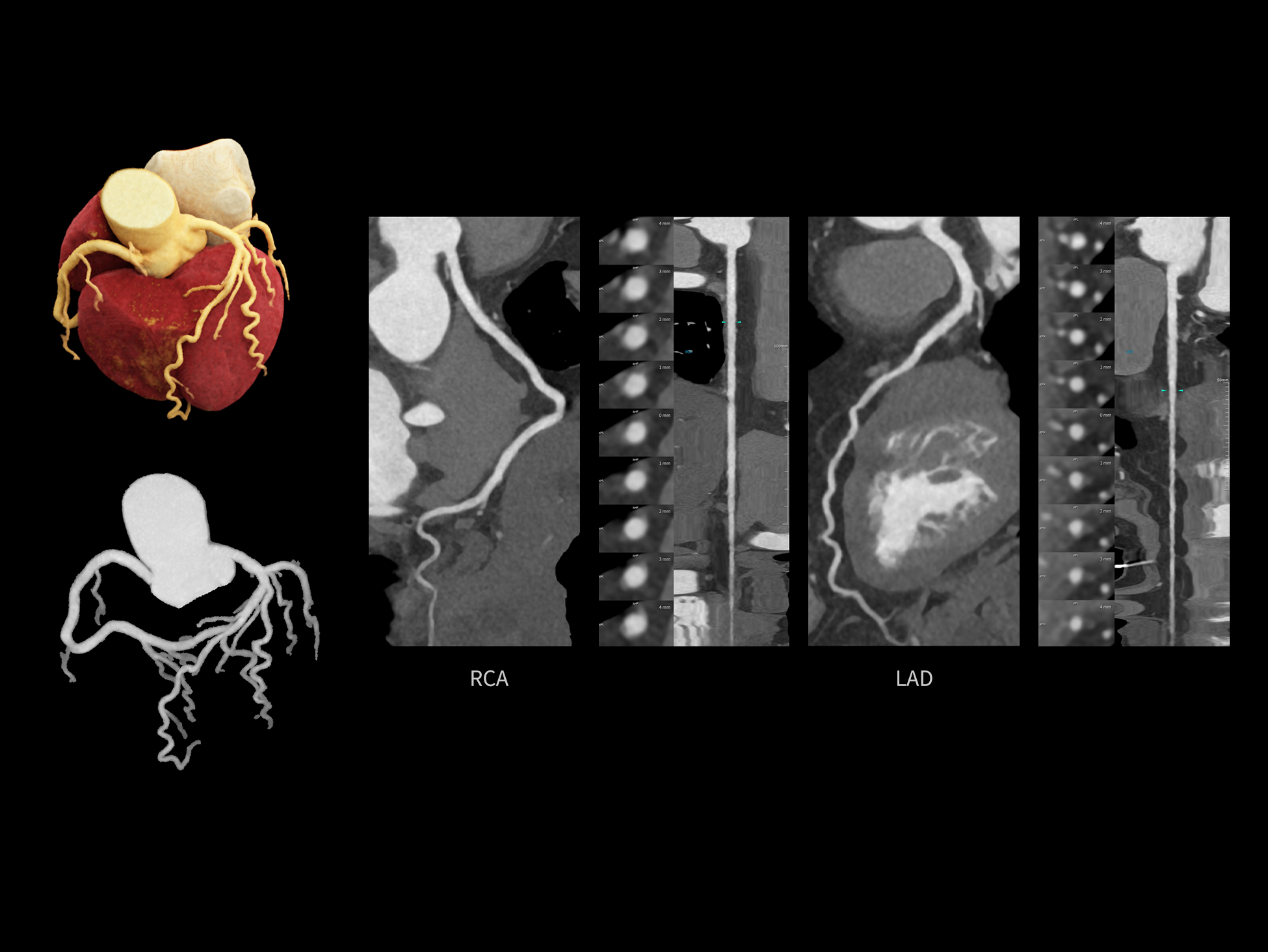
Provides high-quality coronary CTA images across the cardiac cycle, with each phase offering diagnostic-quality visualization to support comprehensive cardiac assessment.

ECG-free Cardiac Static Imaging

Achieves free-breathing, non-ECG-gated coronary CTA with short exposure time, transforming cardiac CT into a low-dose, simplified, and accessible experience.

One-stop Cardiac CTA&CTP* Imaging

Acquires comprehensive cardiac information from a single contrast injection, integrating coronary CTA, myocardial perfusion, and late iodine enhancement for multi-dimensional cardiac assessment.

*CTP analysis is independently registered by United Imaging Intelligence.

Revealing the Whole Body in Spectral Imaging


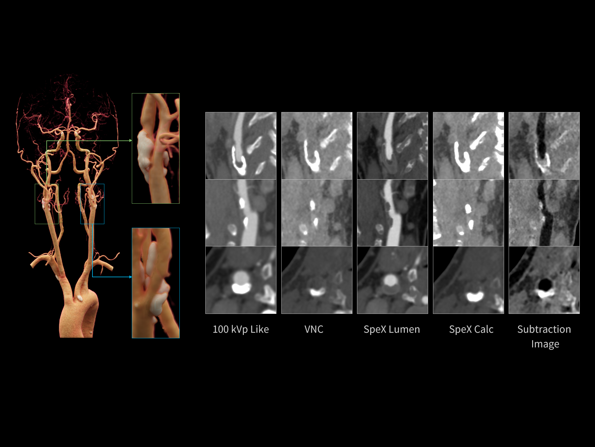
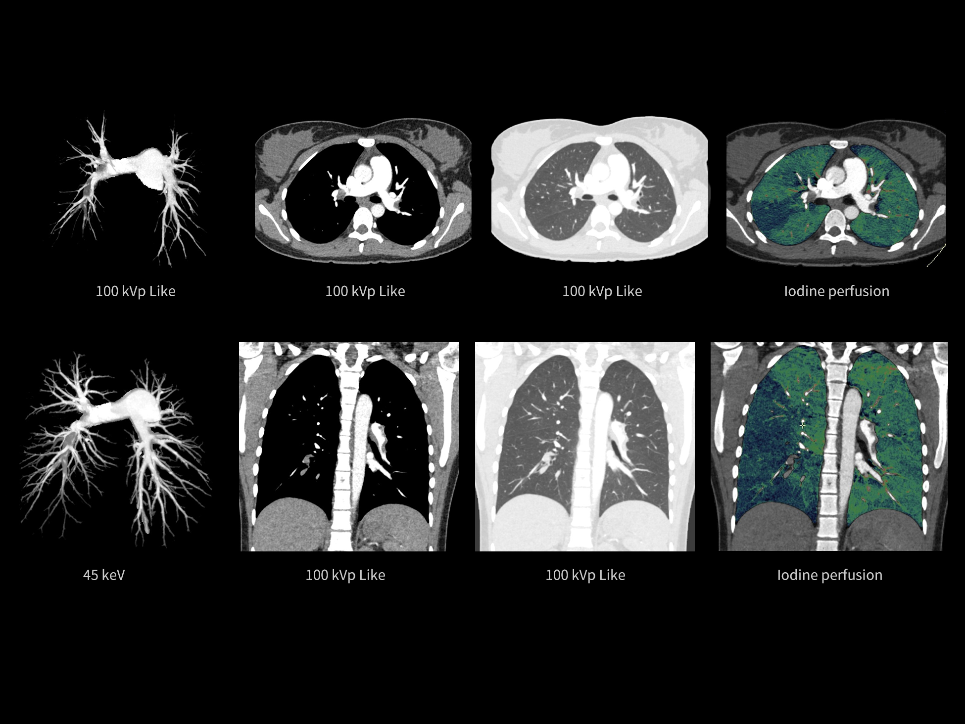
With its dual-wide coverage architecture and advanced spectral imaging capabilities, uCT SiriuX™ extends spectral imaging across a broad range of clinical applications. A maximum spectral imaging field of view of 470 mm, combined with advanced quantitative analysis, supports spectral imaging in diagnosis and research.

Whole-body Spectral Imaging

uCT SiriuX™ features a proprietary dual-wide coverage architecture, enabling 470 mm large field-of-view spectral imaging for full-organ coverage, while seamlessly accommodating patients of varying body types and scan regions. Paired with an spectral reconstruction imaging chain and precise dual-energy data registration, the system effectively suppresses artifacts and boosts signal-to-noise ratio to deliver consistent high-quality spectral imaging across all organs.

470 mm
• Scan FOV of dual-source spectral imaging

SpeX Recon
• AI-powered spectral reconstruction algorithm
Versatile Spectral Analysis Tools

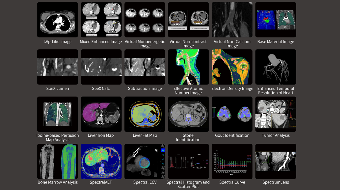
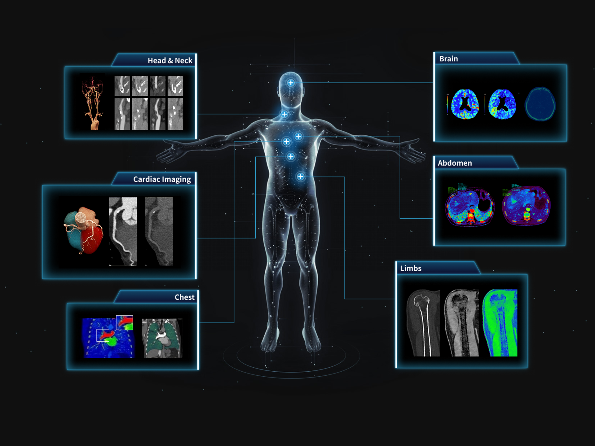
Spectral imaging provides quantitative material-specific and energy-resolved information, with its clinical value further unlocked by an efficient spectral post-processing platform. uCT SiriuX™ offers a comprehensive suite of advanced spectral analysis tools spanning cardiology, oncology, neurology and orthopedics. With a broad range of analytical capabilities and up to 10 basis material pair image types, the platform helps transform spectral data into actionable insights for precision diagnosis and clinical research.

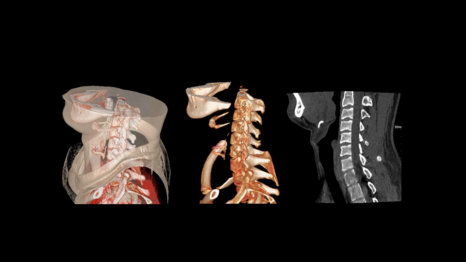
Whole-body Metal Artifact Reduction

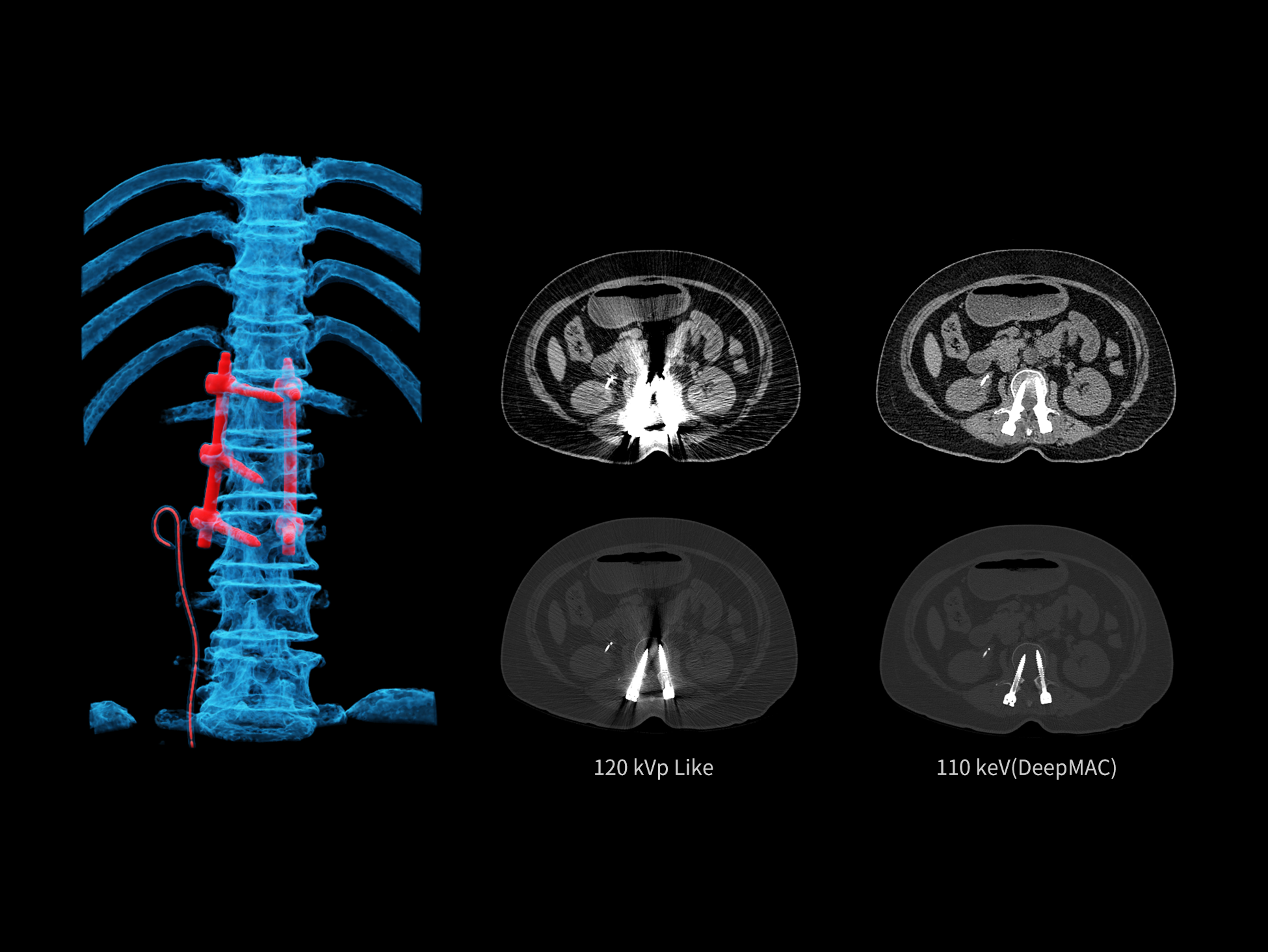
Combining spectral imaging, PureBeam filtration, and DeepMAC deep learning-based metal artifact reduction algorithm, uCT SiriuX™ effectively reduces beam-hardening and metal-induced artifacts, enhancing image quality and diagnostic confidence around metal implants.

Dual-source Spectral Imaging

Dual-source spectral imaging delivers clear high-energy virtual monoenergetic images and efficiently suppresses metal artifacts.

PureBeam – Low-energy photon filtration

Tin Bowtie filter effectively removes low-energy photons, enhancing spectral separation in CT imaging.

DeepMAC – AI-based Metal Artifact Reduction

Powered by a deep learning network model, DeepMAC reduces artifacts from various metal implants, including hip prostheses, spinal fixation devices, orthopedic implants, dental restorations, and endovascular coils, restoring clear anatomical details.

Technical Specifications

Gantry bore size

82 cm

Detectors

2x16 cm Wide-coverage Detectors

Number of slices per rotation

2x640 Slices

Minimum slice thickness

0.5 mm

X-ray tubes

2x 34 MHU Liquid Metal Bearing X-ray Tubes

Rotation time

Up to 0.239 sec/rotation

Temporal resolution

Up to 8 ms with CardioCapture

Table load

Up to 325 kg

kV settings

60/70/80/100/120/140 kV

Generator power

2x100 kW

Disclaimer:

The product may not be commercially available in all countries. Please check with your local United Imaging Healthcare representative for availability.

This product is intended for use by trained medical professionals only. Patients should follow the guidance and instructions provided by healthcare practitioners during operation.

The supported language of the instructions for use is English. Qualified users may access the instruction for use in the appropriate language via the provided link: https://uicloud.com/uclass/docs/en-us/home

Please contact your local United Imaging Healthcare organization for further details.