All Your Needs in One Smart CT
uCT Orion Extra

The uCT Orion Extra integrates AI with CT technology to provide efficient, patient-focused scanning experience and exceptional image clarity. Beyond intelligent scanning, its robust hardware provides the power and reliability to meet diverse clinical needs. As a forward-looking investment, uCT Orion Extra is built for today's demands and tomorrow's value.

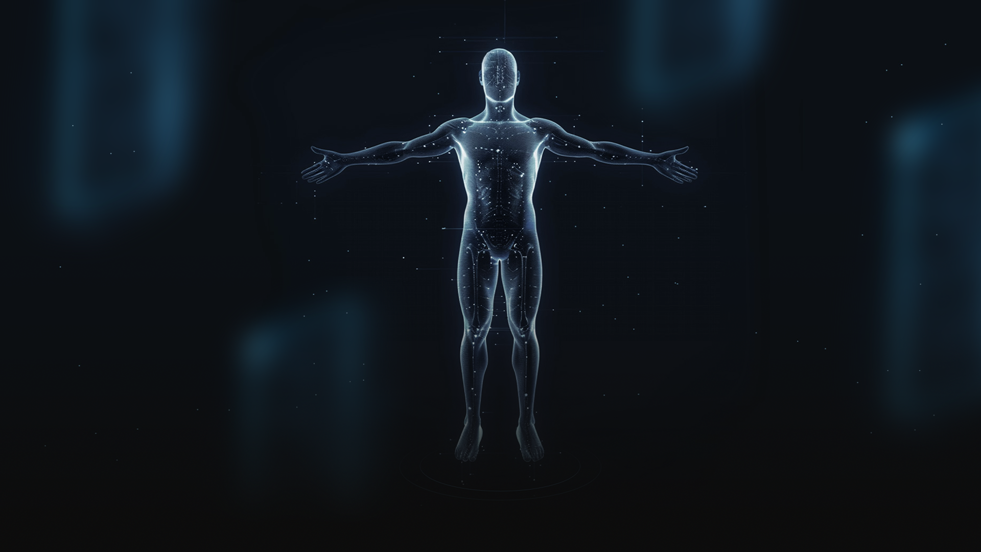
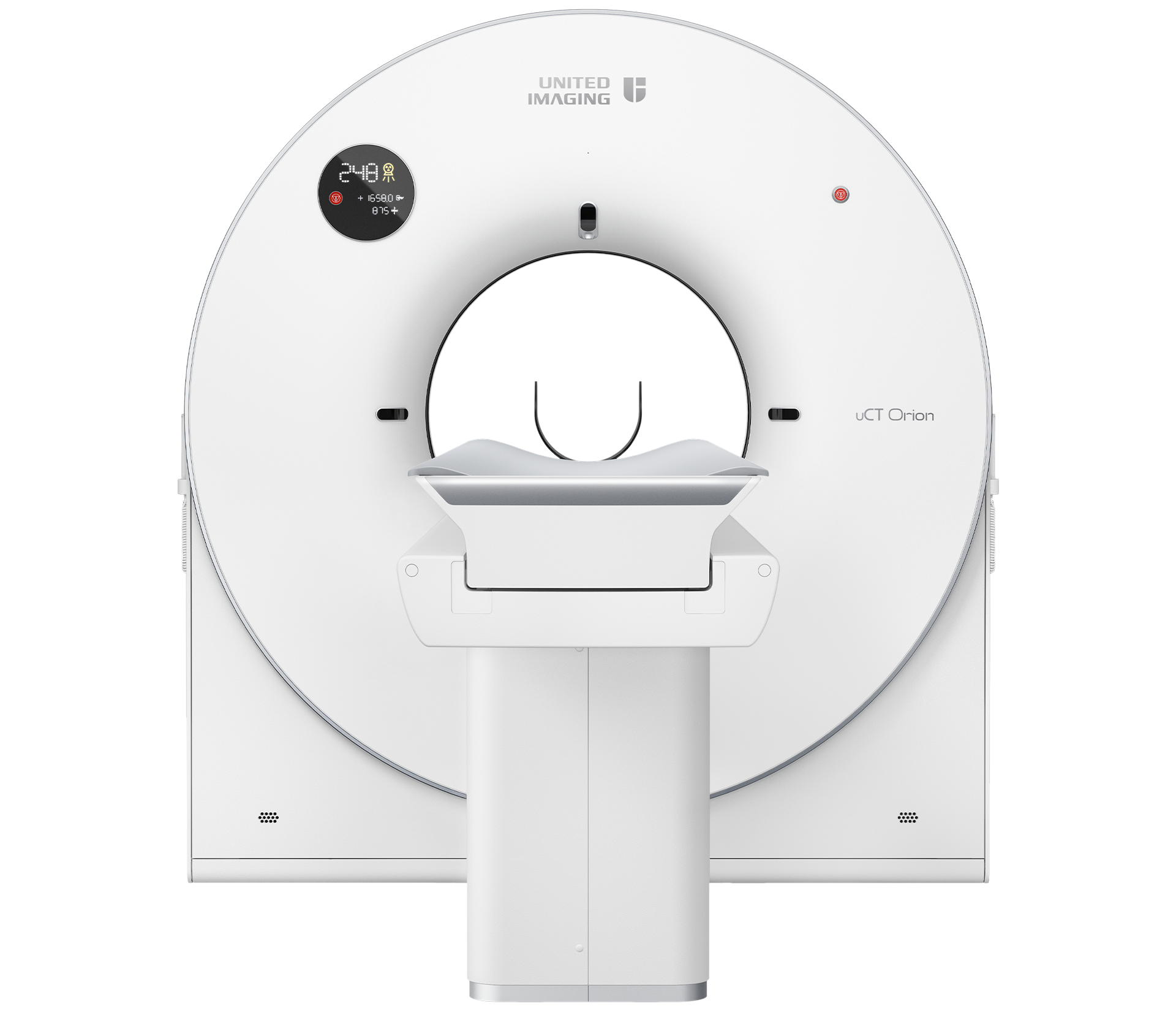
Redefining Workflows with AI


The intelligent workflow of the uCT Orion Extra offers patients and technologists a refreshing scanning experience that far exceeds expectations. With the innovative dual-camera uAI Vision seamlessly combined with multiple AI-powered algorithms, this scanning workflow not only elevates patient care to a next level but also markedly improves scanning efficiency.

uCT Orion Extra Keeps Image Quality on Track

During chest scans, the AI-empowered quality check monitors the patient's breath-hold status. If the patient fails to hold the breath as required, the system triggers real-time prompts, which helps technologists efficiently confirm chest image quality and reduce patient recalls.

Based on AI technology, the uAI Vision in-bore detects patient movement. Once patient's motion is detected, it triggers real - time prompts, enabling technologists to stay vigilant about the patient's scanning status.

It automatically detects non-iatrogenic metal (such as belts, keys, necklaces, etc.) and accurately pinpoints their locations in the scout image. This can remind technologists to remove the metal objects, avoiding the generation of artifacts and enhancing image quality.

Quality Check for Chest

During chest scans, the AI-empowered quality check monitors the patient's breath-hold status. If the patient fails to hold the breath as required, the system triggers real-time prompts, which helps technologists efficiently confirm chest image quality and reduce patient recalls.

Quality Check for Motion

Based on AI technology, the uAI Vision in-bore detects patient movement. Once patient's motion is detected, it triggers real - time prompts, enabling technologists to stay vigilant about the patient's scanning status.

Quality Check for Metal

It automatically detects non-iatrogenic metal (such as belts, keys, necklaces, etc.) and accurately pinpoints their locations in the scout image. This can remind technologists to remove the metal objects, avoiding the generation of artifacts and enhancing image quality.

uCT Orion Extra Guards Every Patient

The uCT Orion Extra's uAI Vision features two cameras, enabling real-time patient monitoring from two distinct views: top-down and close-up. It continuously monitors the patien's status throughout the scan, helping technologists quickly identify issues.

The system uses uAI Vision to proactively identify areas at risk of patient-gantry collisions. Once a potential patient-gantry collision is detected, an anti-collision message will appear before table movement, thereby reducing the likelihood of collisions between the patient and the gantry.

This algorithm is designed for intracerebral hemorrhage (ICH) detection. It provides technologists signals when potential ICH is detected, enabling fast triage of such patient and preserving valuable time for patient assessment.

*Registered by United Imaging Intelligence


uAI Vision for Monitoring

The uCT Orion Extra's uAI Vision features two cameras, enabling real-time patient monitoring from two distinct views: top-down and close-up. It continuously monitors the patien's status throughout the scan, helping technologists quickly identify issues.

Patient-gantry Collision Check

The system uses uAI Vision to proactively identify areas at risk of patient-gantry collisions. Once a potential patient-gantry collision is detected, an anti-collision message will appear before table movement, thereby reducing the likelihood of collisions between the patient and the gantry.

AI-based Brain Hemorrhage Detection*

This algorithm is designed for intracerebral hemorrhage (ICH) detection. It provides technologists signals when potential ICH is detected, enabling fast triage of such patient and preserving valuable time for patient assessment.

*Registered by United Imaging Intelligence


AI-Supported Streamlined Workflow

Based on uAI Vision's detection of 21 anatomical landmarks, the uCT Orion Extra can automatically identify the appropriate scan range based on the selected protocol. The technologist only needs a single click to position the patient for the scout scan, improving preparation efficiency.

The uCT Orion Extra adjust the scan dose based on the patient's body size, scanned region, and examination type. It recommends the appropriate kV and automatically modulates mA during the scan. In addition, AI-based organ recognition enables individualized dose optimization during combined thoracic and abdominal scans. Together, these features achieve optimized dose management and image quality for each patient and scan.

uCT Orion Extra offers a comprehensive suite of console integrated post-processing applications designed to address diverse diagnostic needs across clinical scenarios. These intelligent tools streamline routine workflow by reducing repetitive manual steps, enabling users to complete post-processing more efficiently.

Pre-scan: AI-based Automatic Patient Positioning

Based on uAI Vision's detection of 21 anatomical landmarks, the uCT Orion Extra can automatically identify the appropriate scan range based on the selected protocol. The technologist only needs a single click to position the patient for the scout scan, improving preparation efficiency.

Scan: Intelligent Dose Control

The uCT Orion Extra adjust the scan dose based on the patient's body size, scanned region, and examination type. It recommends the appropriate kV and automatically modulates mA during the scan. In addition, AI-based organ recognition enables individualized dose optimization during combined thoracic and abdominal scans. Together, these features achieve optimized dose management and image quality for each patient and scan.

Post-scan: A Full Suite of Efficient Post-Processing Applications

uCT Orion Extra offers a comprehensive suite of console integrated post-processing applications designed to address diverse diagnostic needs across clinical scenarios. These intelligent tools streamline routine workflow by reducing repetitive manual steps, enabling users to complete post-processing more efficiently.

Boosting Image Quality with AI


AI is transforming CT imaging, and uCT Orion Extra harnesses its power to set a new standard in CT image quality. The DELTA technology elevates image quality across all dimensions , while Motion Freeze suppresses head motion artifacts. These AI-driven algorithms enhance anatomy in every detail, empowering radiologists with greater diagnostic confidence.

DELTA*- Deep Learning Trained Algorithm

Unlike conventional 2D networks that process only one image at a time, 3D neural networks can handle volumetric data inputs. This capability preserves data continuity and enhances reconstruction speed, accuracy and reliability.

Learn more

The network learns by comparing noisy inputs with higher-quality references. Trained on over one million cases from diverse clinical scenarios, it identifies and suppress noise while preserving anatomical detail.

The network is pushed beyond routine — tested with rare, and extreme cases that mirror real-world clinical complexity.

*Optional

Specialized 3D network design

Unlike conventional 2D networks that process only one image at a time, 3D neural networks can handle volumetric data inputs. This capability preserves data continuity and enhances reconstruction speed, accuracy and reliability.

Learn more

Millions-scale datasets for training

The network learns by comparing noisy inputs with higher-quality references. Trained on over one million cases from diverse clinical scenarios, it identifies and suppress noise while preserving anatomical detail.

Promote reliability and robustness

The network is pushed beyond routine — tested with rare, and extreme cases that mirror real-world clinical complexity.

*Optional
DELTA elevates image quality across all dimensions
In contrast to conventional methods such as Filtered Back-Projection (FBP) and Model-Based Iterative Reconstruction (MBIR), DELTA attains a balance among high image quality, low radiation dose, and fast reconstruction speed - a balance that has long been deemed challenging to achieve. With this achievement, DELTA sets a new benchmark in CT imaging.
163%*

Up to 163% LCD improvement at the same dose.

74%*

Up to 74% spatial resolution improvement at the same dose.

80%*

Up to 80% radiation dose reduction at the same low contrast detectability (LCD).

98%*

Up to 98% image noise reduction at the same dose.


*The above data are based on phantom tests comparing DELTA with FBP

DELTA reduces image noise and enhances low contrast detectability
  • Scan Parameter
  • kV: 120
    mAs: 285
    CTDIvol: 45.8 mGy
    Eff.Dose: 1.7 mSv
  • Recon Parameter
  • Matrix: 512 * 512
    Slice: 1.0 * 0.8 mm
    HIR: H_SOFT_B
    DL-Denoise level: 3
    WW/WL: 80/35
  • Contrast
DELTA: High Image Quality at Low Radiation Dose
  • Scan Parameter
  • kV: 60
    mAs: 210
    CTDIvol: 1.8 mGy
    Eff.Dose: 0.8 mSv
  • Recon Parameter
  • Matrix: 512 * 512
    Slice: 1.0 * 0.5 mm
    HIR: B_SHARP
    DL-Denoise level: 3
    WW/WL: 1500/-500
  • Contrast
DELTA optimizes image quality in CT angiography
  • Scan Parameter
  • kV: 100
    mAs: 130
    CTDIvol: 7.568 mGy
    Eff.Dose: 0.54mSv
  • Recon Parameter
  • Matrix: 512 * 512
    Slice: 1.0 * 0.5 mm
    HIR: H_SOFT+
    DL-Denoise level: 3
    WW/WL: 820/250
  • Contrast
  • 350mgl/ml
    65ml
    4.2ml/s
Motion Freeze AI for the Suppression of Head Motion Artifacts

Patient head movement typically involves multiple motion patterns, which makes it challenging to obtain large datasets of head motion artifacts for training deep learning algorithms. To construct the training dataset, the Motion Freeze algorithm generates simulated artifacts on gold-standard images along the X, Y, and Z axes, encompassing rotation, translation, oscillation, and mixed motion patterns. By introducing diverse motion artifacts, this approach enables the trained network model to handle a broad spectrum of motion conditions.

Learn more

Motion Freeze effectively suppresses head motion artifacts, providing a clear view of brain structures without obscuring lesions. By minimizing the need for repeated scans, it saves time, conserves resources, and avoids unnecessary radiation exposure for patients.

3D modeling of the motion pattern to restore the real clinical situation

Patient head movement typically involves multiple motion patterns, which makes it challenging to obtain large datasets of head motion artifacts for training deep learning algorithms. To construct the training dataset, the Motion Freeze algorithm generates simulated artifacts on gold-standard images along the X, Y, and Z axes, encompassing rotation, translation, oscillation, and mixed motion patterns. By introducing diverse motion artifacts, this approach enables the trained network model to handle a broad spectrum of motion conditions.

Learn more

Head motion artifact suppression

Motion Freeze effectively suppresses head motion artifacts, providing a clear view of brain structures without obscuring lesions. By minimizing the need for repeated scans, it saves time, conserves resources, and avoids unnecessary radiation exposure for patients.

Exceptional Hardware Meets Your Needs


The uCT Orion Extra is built on a powerful and reliable imaging chain, supported by advanced hardware that allows even complex clinical scans to be performed with ease. This robust system delivers high-quality images, providing clinicians with clear anatomical details and reinforcing confidence in diagnosis.

Technical Details

Powerful and reliable imaging chain

A Smart Investment for Long - Term Value


The uCT Orion Extra is designed to optimize cost efficiency across the entire lifecycle. Its compact footprint simplifies installation and reduces upfront facility expenses, while uEco Mode lowers daily power consumption through sustainable design. uRemote Assistance and Remote Update help users reduce downtime, making daily scanning and operations smoothly. With the uCT Orion Extra, every aspect of your investment is optimized to deliver lasting value—today and for years to come.