Cardiac CT is essential for non-invasive coronary artery assessment, but it remains inherently challenged by motion artifacts, as heart movement varies with patient conditions. Although hardware advances—such as faster rotation speeds and wider detector coverage—have improved image quality, limitations persist, particularly in cases of high or irregular heart rates. These challenges highlight the need for a smarter approach that extends beyond hardware.
The integration of artificial intelligence offers a practical advancement, helping CT systems overcome motion-related limitations and realize their full diagnostic potential. CardioCapture, an AI-powered algorithm, actively corrects motion artifacts, delivering consistent image quality and improving clinical efficiency.

From Hardware to Intelligence: CardioCapture Tackles Residual Motion in Coronary CT Imaging

CardioCapture is an AI-empowered algorithm designed to address these challenges through intelligent motion correction. CardioCapture reconstructs coronary images from multiple cardiac phases near the reference phase to analyze vessel motion and generate a motion map of the vessels, which is then used to correct motion artifacts in the reference phase image. At the core of this process is precise motion trajectory estimation of the coronary arteries. CardioCapture uses deep learning to automatically extract coronary artery centerlines. Even under severe motion artifacts caused by high heart rates, centerlines remain the most reliable representation of vessel location. By leveraging AI for this step, the algorithm provides consistent, high-quality imaging under challenging conditions.

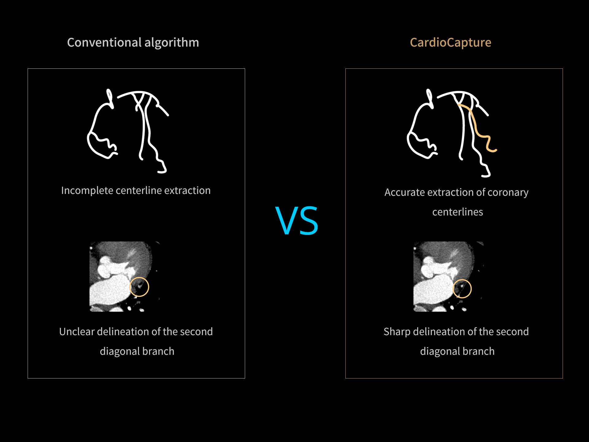
Capture more details with precise AI extraction

Conventional vessel extraction methods typically rely on CT value thresholds and fixed coronary models, which often fail, particularly when dealing with vessels affected by motion artifacts. In contrast, CardioCapture excels at accurately extracting the centerlines of various types of coronary arteries, even in challenging cases involving poor vessel quality or distal vessels.

Improve the effective temporal resolution to 25ms

With the AI-based coronary artery motion correction technology, our CT scanner is able to break the limit of system native temporal resolution and achieve an effective temporal resolution of 25ms, which greatly enhances the success rate and image clarity of coronary CTA imaging.

Fully integrated on console

CardioCapture is built into the reconstruction process of coronary CTA protocols and corrected images can be directly generated on the console after each cardiac scan. The time consumed for data transfer between the console and workstation can now be fully eliminated.

The use of MCA(CardioCapture) significantly enhanced image quality and diagnostic performance in cardiac CT. Subjective image quality improved markedly, with vessel interpretability increasing from 89.9 to 98.8%. Diagnostic accuracy also saw substantial gains: the area under the curve (AUC) improved from 0.58 to 0.83 in patient-based analysis (p = 0.04), and from 0.81 to 0.92 in vessel-based analysis (p < 0.001).

< Improving Image Quality and Diagnostic Performance of CCTA in Patients with Challenging Heart Rate Conditions using a Deep Learning-based Motion Correction Algorithm >.

—《 Curr Med Imaging. 2024;20:e15734056315753. 》


In this study of 192 patients undergoing CCTA, a deep learning-based motion correction algorithm (MCA) was evaluated for its effectiveness across different heart rates. Patients were divided into two groups: those with HR <75 bpm (n=82) and HR ≥75 bpm (n=110). The MCA significantly improved subjective image quality in the higher HR group (Group 2) across all evaluated metrics—vessel visualization, sharpness, diagnostic confidence, and overall image quality. The MCA effectively enhances image quality and diagnostic confidence in patients with elevated heart rates, enabling consistent, interpretable CCTA results regardless of HR.

< Deep Learning–Based Motion Correction in Projection Domain for Coronary Computed Tomography Angiography: A Clinical Evaluation >.

—《Journal of Computer Assisted Tomography 47(6):p 898-905, 11/12 2023.》


In 90 patients with heart rates over 75 bpm, AI-assisted motion correction significantly enhanced coronary segment image quality in CCTA exams. Compared to images reconstructed with automatic phase selection alone, the addition of motion correction improved the average quality rating from 3.64 ± 0.55 to 3.85 ± 0.37. This gain was achieved without manual radiologist involvement, demonstrating that AI-based correction—combined with 0.25-second rotation speed and 16-cm z-coverage—enables efficient, high-quality image reconstruction even under challenging heart rate conditions.

< Image quality of automatic coronary CT angiography reconstruction for patients with HR ≥ 75 bpm using an AI-assisted 16-cm z-coverage CT scanner >.

—《BMC Med Imaging. 2021 Feb 11;21(1):24..》


The AI-assisted automatic reconstruction achieved a higher average Likert score (3.48 ± 0.62) compared to manually reconstructed images (3.32 ± 0.67, P < 0.001). These results indicate that a CT scanner with 0.25-second rotation speed, 16 cm coverage, and integrated AI-based motion correction and auto-phase selection can outperform manual reconstruction, suggesting a more efficient workflow with reduced reliance on radiologist input.

< Automatic vs manual coronary CT angiography reconstruction for whole-heart coverage CT scanner: a comparison study in general patient population >.

—《 Journal of X-Ray Science and Technology. 2022;30(2):389-398. 》


The motion correction algorithm (MCA) significantly improves image quality and diagnostic confidence at both optimal and nonoptimal phases, maintaining reliable coronary assessment within a 4% phase deviation. The deep learning-based MCA enables dependable morphological and functional CCTA evaluation even in patients with high heart rates, allowing for slight phase deviations without compromising diagnostic reliability.

< Deep learning-based motion correction algorithm for coronary CT angiography: Lowering the phase requirement for morphological and functional evaluation >.

—《Journal of Applied Clinical Medical Physics. 》